Copyright
©The Author(s) 2004.
World J Gastroenterol. Jan 1, 2004; 10(1): 17-21
Published online Jan 1, 2004. doi: 10.3748/wjg.v10.i1.17
Published online Jan 1, 2004. doi: 10.3748/wjg.v10.i1.17
Figure 1 Antral biopsy showing focal intestinal metaplasia.
This composite figure shows the H&E appearance, negative p16 staining (with some positive inflammatory cells in the background) and positive pRb staining limited to the neck region of the glands. Original magnification 200 × .
Figure 2 Gastric antral carcinoma of intestinal type.
The same 0.6 mm TMA core is shown stained with H&E, p16 and pRb. The latter is negative whereas p16 shows strong nuclear and cytoplasmic staining. The cytoplasmic staining was ignored for the scoring. Original magnification 200 × .
Figure 3 Gastric antral carcinoma of intestinal type.
The same 0.6 mm TMA core is shown stained with H&E, p16 and pRb. The latter shows strong and crisp nuclear staining whereas p16 is negative. Original magnification 200 × .
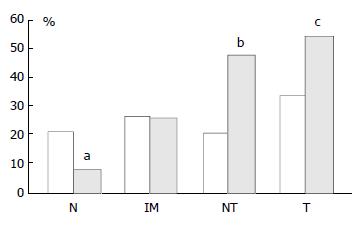
Figure 4 Graph showing percentage of p16 positive cases as pairs of proximal (empty columns, left hand sides) and distal (black columns, right hand sides) tissue samples.
N: Biopsies showing normal mucosa, IM: Biopsies showing intestinal metaplasia, NT: Non-involved mucosa from gastric cancer resection specimens, T: Tumour from gastric cancer resection specimens. There was a significant stepwise increase in expres-sion from normal mucosa→intestinal metaplasia→non-involved mucosa from cancer resections→carcinoma in the distal stomach only. aThere was a significantly lower p16 ex-pression in distal normal mucosa than in proximal normal mucosa, P = 0.0045. b and c: There was a significantly higher p16 expression in both non-involved as well as carcinoma from cancer resections from distal compared with proximal stomach, bP = 0.0048 and cP = 0.036.
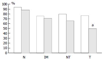
Figure 5 Graph showing percentage of pRb positive cases as pairs of proximal (empty columns, left hand side) and distal (black columns, right hand side) tissue samples.
N: Biopsies showing normal mucosa, IM: Biopsies showing intestinal metaplasia, NT: Non-involved mucosa from gastric cancer re-section specimens, T: Tumour from gastric cancer resection specimens. There was a significant stepwise decrease in ex-pression from normal mucosa→intestinal metaplasia→non-in-volved mucosa from cancer resections→carcinoma in both the distal and proximal stomach although in the latter location it was most likely due to the high expression in normal mucosa compared with the other types of tissues. aThere was a signifi-cantly lower pRb expression in distal than in proximal carcinomas, P = 0.0047.
- Citation: Gulmann C, Hegarty H, Grace A, Leader M, Patchett S, Kay E. Differences in proximal (cardia) versus distal (antral) gastric carcinogenesis via retinoblastoma pathway. World J Gastroenterol 2004; 10(1): 17-21
- URL: https://www.wjgnet.com/1007-9327/full/v10/i1/17.htm
- DOI: https://dx.doi.org/10.3748/wjg.v10.i1.17


