Copyright
©The Author(s) 2004.
World J Gastroenterol. Jan 1, 2004; 10(1): 132-135
Published online Jan 1, 2004. doi: 10.3748/wjg.v10.i1.132
Published online Jan 1, 2004. doi: 10.3748/wjg.v10.i1.132
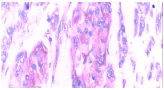
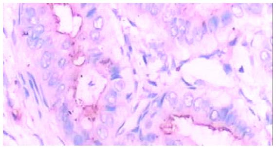
Figure 1 Ductal adenocarcinoma in pancreatic head (Immunohisto-chemistry LSAB method, × 400).
Brown yellow staining of SST2R gene expression in cell membrane/cytoplasm.
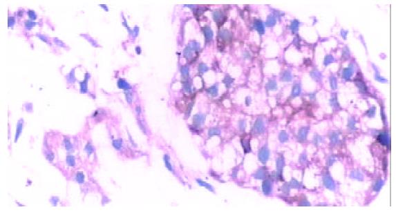
Figure 2 Pancreatic ductal adenocarcinoma in pancreatic body and tail (Immunohistochemistry LSAB method, × 400).
Brown yellow staining of SST2R gene expression in cell membrane/ cytoplasm.
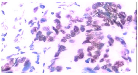
Figure 3 Ductal adenocarcinoma in pancreatic head (EnVisionTM method, × 400).
Brown yellow staining of p53 gene expression in cell nucleus.
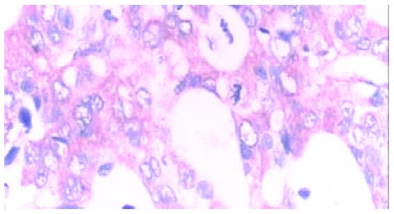
Figure 4 Ductal adenocarcinoma in pancreatic body and tail (EnVisionTM method, × 400).
Brown yellow staining of ras gene expression located in cytoplasm.
Figure 5 Ductal adenocarcinoma in pancreatic body and tail (EnVisionTM method, × 400).
Brown yellow staining of DPC4 gene expression located in cytoplasm.
- Citation: Qin RY, Fang RL, Gupta MK, Liu ZR, Wang DY, Chang Q, Chen YB. Alteration of somatostatin receptor subtype 2 gene expression in pancreatic tumor angiogenesis. World J Gastroenterol 2004; 10(1): 132-135
- URL: https://www.wjgnet.com/1007-9327/full/v10/i1/132.htm
- DOI: https://dx.doi.org/10.3748/wjg.v10.i1.132


