Copyright
©The Author(s) 2017.
World J Meta-Anal. Dec 26, 2017; 5(6): 132-149
Published online Dec 26, 2017. doi: 10.13105/wjma.v5.i6.132
Published online Dec 26, 2017. doi: 10.13105/wjma.v5.i6.132
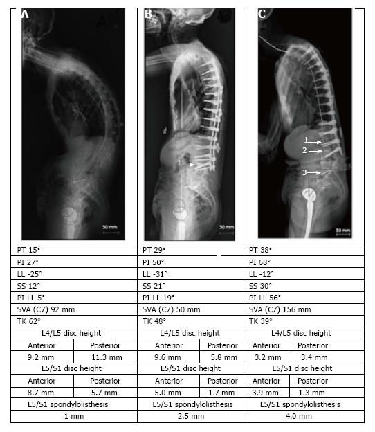
Figure 2 Example of distal segment degeneration/ failure.
This event was observed after surgical correction of adult spine deformity in a patient (67 years old male) with idiopathic kyphoscoliosis complicated by degenerative disc disease and lumbar stenosis. A: Severe preoperative spinal kyphosis; B: Postoperative correction by T3-L4 posterior instrumented fusion with L3/L4 decompression, transforaminal interbody fusion by cage placement (1), and multilevel (T5-T11) Smith-Petersen osteotomy; C: Distal segment degeneration/ failure at 35 mo follow-up with deformation and/or fracture of L3 (1) subsidence of the interbody cage (2), collapse of L4/L5 and L5/S1 intervertebral discs with L5/S1 spondylolisthesis (3), loss of sagittal balance, and progression of proximal junctional kyphosis. Interestingly, these changes were accompanied by significant increase of pelvic incidence (PI) from 27° to 68° due to simultaneous increase of pelvic tilt (PT) and sacral slope which suggested that position of sacrum relatively to pelvis changed after surgery. Likely, this is the result of displacement in the sacroiliac joints. This finding contravenes the concept concerning postoperative stability of PI.
- Citation: Barton C, Noshchenko A, Patel VV, Cain CMJ, Kleck C, Burger EL. Different types of mechanical complications after surgical correction of adult spine deformity with osteotomy. World J Meta-Anal 2017; 5(6): 132-149
- URL: https://www.wjgnet.com/2308-3840/full/v5/i6/132.htm
- DOI: https://dx.doi.org/10.13105/wjma.v5.i6.132


