
















©The Author(s) 2024.
World J Meta-Anal. Sep 18, 2024; 12(3): 94519
Published online Sep 18, 2024. doi: 10.13105/wjma.v12.i3.94519
Published online Sep 18, 2024. doi: 10.13105/wjma.v12.i3.94519
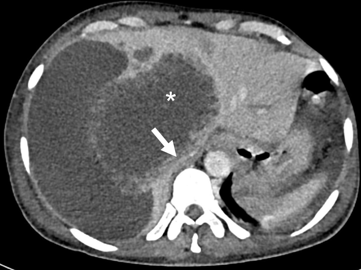
Figure 2 Axial contrast-enhanced computed tomography image showing a large ruptured amebic liver abscess (asterisk) in the caudate lobe, exerting pressure on the inferior vena cava (arrow).
Note the fluid collection in perihepatic space resulting from the ruptured abscess.
- Citation: Arya R, Kumar R, Priyadarshi RN, Narayan R, Anand U. Vascular complications of liver abscess: A literature review. World J Meta-Anal 2024; 12(3): 94519
- URL: https://www.wjgnet.com/2308-3840/full/v12/i3/94519.htm
- DOI: https://dx.doi.org/10.13105/wjma.v12.i3.94519