Copyright
©The Author(s) 2021.
World J Clin Cases. Mar 6, 2021; 9(7): 1668-1675
Published online Mar 6, 2021. doi: 10.12998/wjcc.v9.i7.1668
Published online Mar 6, 2021. doi: 10.12998/wjcc.v9.i7.1668
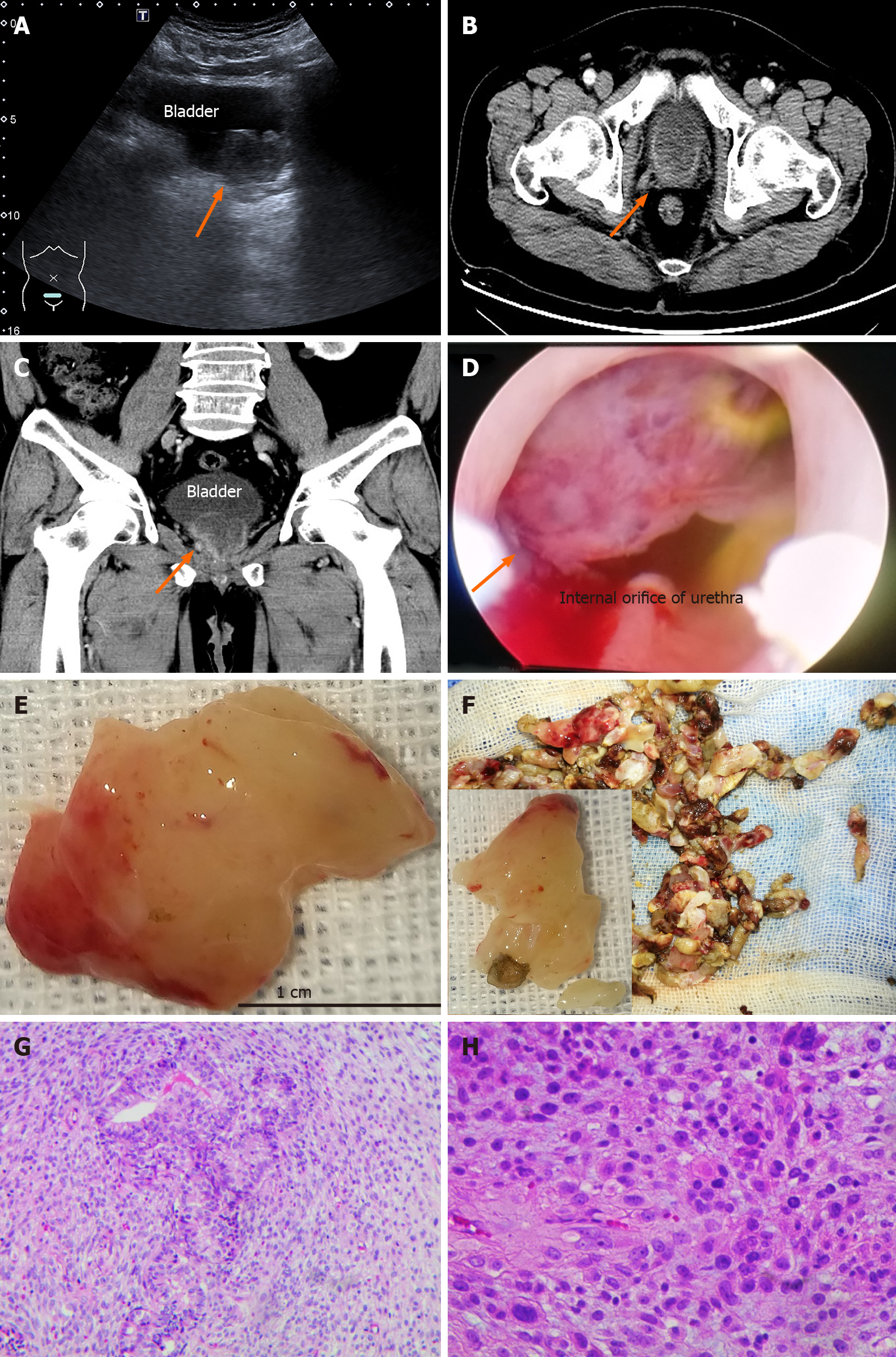
Figure 1 Clinical and pathological features of case 2 at the diagnosis of sarcomatoid carcinoma.
A: Ultrasound: The size of the prostate was 3.9 cm × 3.2 cm, and a 1.5 cm × 1.1 cm round mass was present in the gland (arrow); B and C: The mass was approximately 4.1 cm × 3.0 cm × 4.0 cm on computed tomography (arrow), and enhanced scanning was uneven. No obvious abnormality was found in the bilateral seminal vesicles; D-F: A white, narrow pedicled, spherical solid tumor blocked the internal orifice of the urethra. The spherical lesion mainly arose from the 8-11 o'clock position of the prostate and resembled fish flesh in sections (arrow); G and H: Various heteromorphic tumor cells showed infiltrating growth, which included immature small round cells, subepithelial cells, spindle cells, lipoblasts and tumor giant cells (arrow), etc. G: Original magnification × 100; H: × 200.
- Citation: Wei W, Li QG, Long X, Hu GH, He HJ, Huang YB, Yi XL. Sarcomatoid carcinoma of the prostate with bladder invasion shortly after androgen deprivation: Two case reports. World J Clin Cases 2021; 9(7): 1668-1675
- URL: https://www.wjgnet.com/2307-8960/full/v9/i7/1668.htm
- DOI: https://dx.doi.org/10.12998/wjcc.v9.i7.1668


