
















©The Author(s) 2021.
World J Clin Cases. Dec 26, 2021; 9(36): 11495-11503
Published online Dec 26, 2021. doi: 10.12998/wjcc.v9.i36.11495
Published online Dec 26, 2021. doi: 10.12998/wjcc.v9.i36.11495
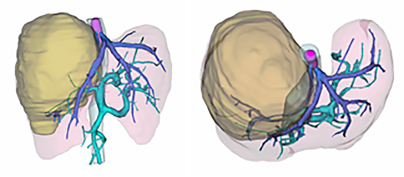
Figure 2 The three-dimensional reconstruction of this patient was performed by using computed tomography scanning images.
The tumor and thrombus are shown in yellow and pink, while the hepatic vein and portal vein are shown in deep and light blue, respectively.
- Citation: Zhang ZY, Zhang EL, Zhang BX, Zhang W. Surgery for hepatocellular carcinoma with tumor thrombosis in inferior vena cava: A case report. World J Clin Cases 2021; 9(36): 11495-11503
- URL: https://www.wjgnet.com/2307-8960/full/v9/i36/11495.htm
- DOI: https://dx.doi.org/10.12998/wjcc.v9.i36.11495