Copyright
©The Author(s) 2021.
World J Clin Cases. Dec 26, 2021; 9(36): 11495-11503
Published online Dec 26, 2021. doi: 10.12998/wjcc.v9.i36.11495
Published online Dec 26, 2021. doi: 10.12998/wjcc.v9.i36.11495
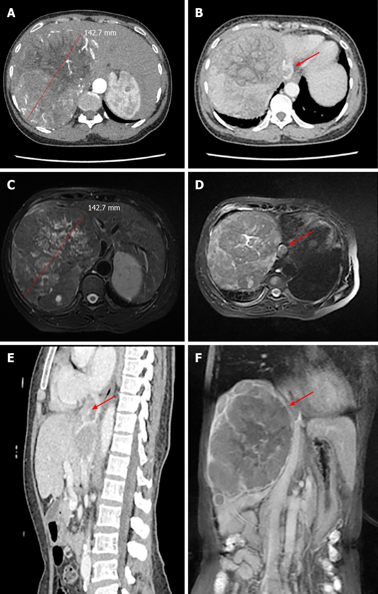
Figure 1 Preoperative imaging studies.
A: Liver-enhanced computed tomography (CT) showing the diameter of the tumor lesion in the liver; B: The tumor thrombus was detected in the supra-hepatic inferior vena cava (red arrow); C: Magnetic resonance imaging (MRI) showing the diameter of the tumor lesion in the liver; D: The tumor thrombus was detected in the supra-hepatic inferior vena cava (red arrow); E and F: The sagittal plane was reconstructed by CT (E) and MRI (F) and shows the position of the tumor thrombus (red arrow).
- Citation: Zhang ZY, Zhang EL, Zhang BX, Zhang W. Surgery for hepatocellular carcinoma with tumor thrombosis in inferior vena cava: A case report. World J Clin Cases 2021; 9(36): 11495-11503
- URL: https://www.wjgnet.com/2307-8960/full/v9/i36/11495.htm
- DOI: https://dx.doi.org/10.12998/wjcc.v9.i36.11495


