Copyright
©The Author(s) 2021.
World J Clin Cases. Dec 26, 2021; 9(36): 11467-11474
Published online Dec 26, 2021. doi: 10.12998/wjcc.v9.i36.11467
Published online Dec 26, 2021. doi: 10.12998/wjcc.v9.i36.11467
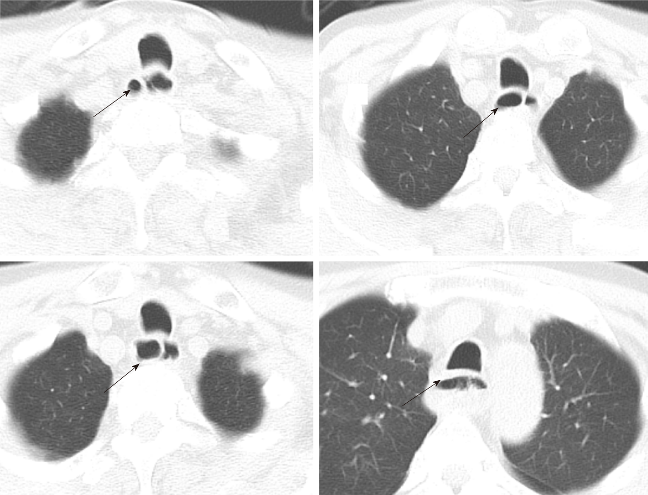
Figure 2 Computed tomography of the esophagus before endoscopic treatment in this patient.
The esophageal cavity was dilated with an irregular diaphragmatic shadow, linear-enhancement is observed in the mucosa after contrast enhancement and some segments of the esophagus have a “double-barreled” appearance. The arrow indicates a “double-barreled” appearance on computed tomography scan.
- Citation: Hu JW, Zhao Q, Hu CY, Wu J, Lv XY, Jin XH. Rare spontaneous extensive annular intramural esophageal dissection with endoscopic treatment: A case report. World J Clin Cases 2021; 9(36): 11467-11474
- URL: https://www.wjgnet.com/2307-8960/full/v9/i36/11467.htm
- DOI: https://dx.doi.org/10.12998/wjcc.v9.i36.11467


