Copyright
©The Author(s) 2021.
World J Clin Cases. Dec 26, 2021; 9(36): 11382-11391
Published online Dec 26, 2021. doi: 10.12998/wjcc.v9.i36.11382
Published online Dec 26, 2021. doi: 10.12998/wjcc.v9.i36.11382
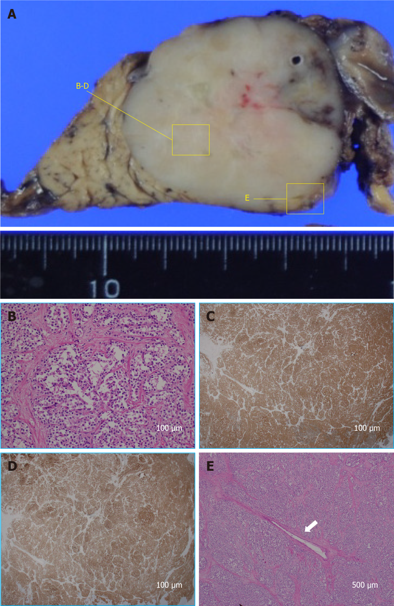
Figure 7 Hematoxylin and eosin staining and pathological findings.
A: Cut surface of the resected specimen is a white solid mass separate from the pancreas; B: Histologically, hematoxylin and eosin staining (× 200) shows a pseudopapillary structure and a pseudorosette structure like the biopsy sample obtained by endoscopic ultrasound-guided fine needle; C and D: Immunostaining also shows that the tumor is β-catenin-positive (C, × 20) and CD10-positive (D, × 20). The pathological findings result in the final diagnosis of SPN. The main pancreatic duct is very narrow; E: Histologically, there is no malignant infiltration, and the tumor surrounds the main pancreatic duct, compressing it from all directions (× 40).
- Citation: Nakashima S, Sato Y, Imamura T, Hattori D, Tamura T, Koyama R, Sato J, Kobayashi Y, Hashimoto M. Solid pseudopapillary neoplasm of the pancreas in a young male with main pancreatic duct dilatation: A case report. World J Clin Cases 2021; 9(36): 11382-11391
- URL: https://www.wjgnet.com/2307-8960/full/v9/i36/11382.htm
- DOI: https://dx.doi.org/10.12998/wjcc.v9.i36.11382


