Copyright
©The Author(s) 2021.
World J Clin Cases. Dec 26, 2021; 9(36): 11382-11391
Published online Dec 26, 2021. doi: 10.12998/wjcc.v9.i36.11382
Published online Dec 26, 2021. doi: 10.12998/wjcc.v9.i36.11382
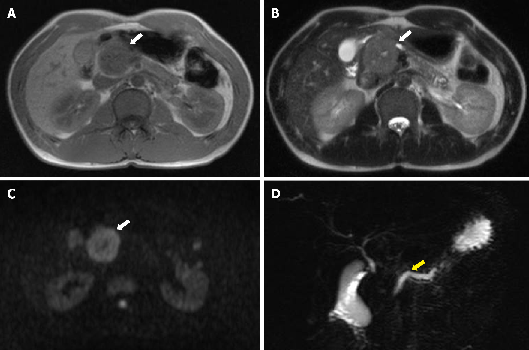
Figure 3 Magnetic resonance imaging and magnetic resonance cholangiopancreatography.
A-C: Magnetic resonance imaging shows low intensity on T1- and T2-weighted images and high intensity on diffusion-weighted images (white arrows); D: Magnetic resonance cholangiopancreatography shows dilatation of the main pancreatic duct upstream of the mass (yellow arrow).
- Citation: Nakashima S, Sato Y, Imamura T, Hattori D, Tamura T, Koyama R, Sato J, Kobayashi Y, Hashimoto M. Solid pseudopapillary neoplasm of the pancreas in a young male with main pancreatic duct dilatation: A case report. World J Clin Cases 2021; 9(36): 11382-11391
- URL: https://www.wjgnet.com/2307-8960/full/v9/i36/11382.htm
- DOI: https://dx.doi.org/10.12998/wjcc.v9.i36.11382


