Copyright
©The Author(s) 2021.
World J Clin Cases. Nov 16, 2021; 9(32): 9954-9959
Published online Nov 16, 2021. doi: 10.12998/wjcc.v9.i32.9954
Published online Nov 16, 2021. doi: 10.12998/wjcc.v9.i32.9954
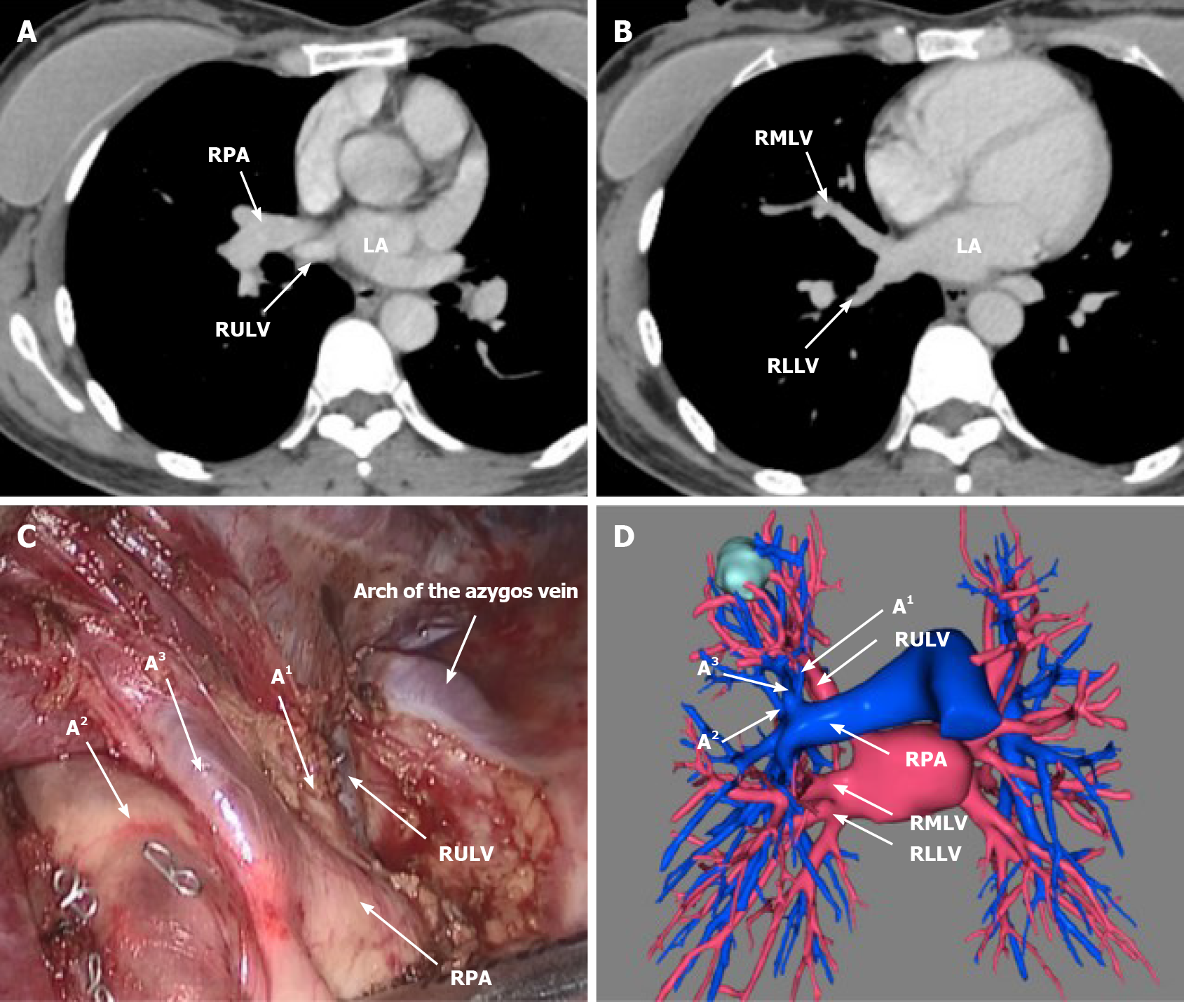
Figure 2 Anatomic aberration of pulmonary vessels.
A: Right upper lobe vein (RULV) lies behind right pulmonary artery (RPA) in thin-section computed tomography (CT); 2B: Right middle lobe vein joins right lower lobe vein to form one common trunk vein drained into left atrium in thin-section CT; C: RULV lies behind right pulmonary artery under thoracoscope; D: Three-dimensional reconstruction of pulmonary vessels.
- Citation: Wang FQ, Zhang R, Zhang HL, Mo YH, Zheng Y, Qiu GH, Wang Y. Rare location and drainage pattern of right pulmonary veins and aberrant right upper lobe bronchial branch: A case report. World J Clin Cases 2021; 9(32): 9954-9959
- URL: https://www.wjgnet.com/2307-8960/full/v9/i32/9954.htm
- DOI: https://dx.doi.org/10.12998/wjcc.v9.i32.9954


