Copyright
©The Author(s) 2021.
World J Clin Cases. Sep 26, 2021; 9(27): 8186-8191
Published online Sep 26, 2021. doi: 10.12998/wjcc.v9.i27.8186
Published online Sep 26, 2021. doi: 10.12998/wjcc.v9.i27.8186
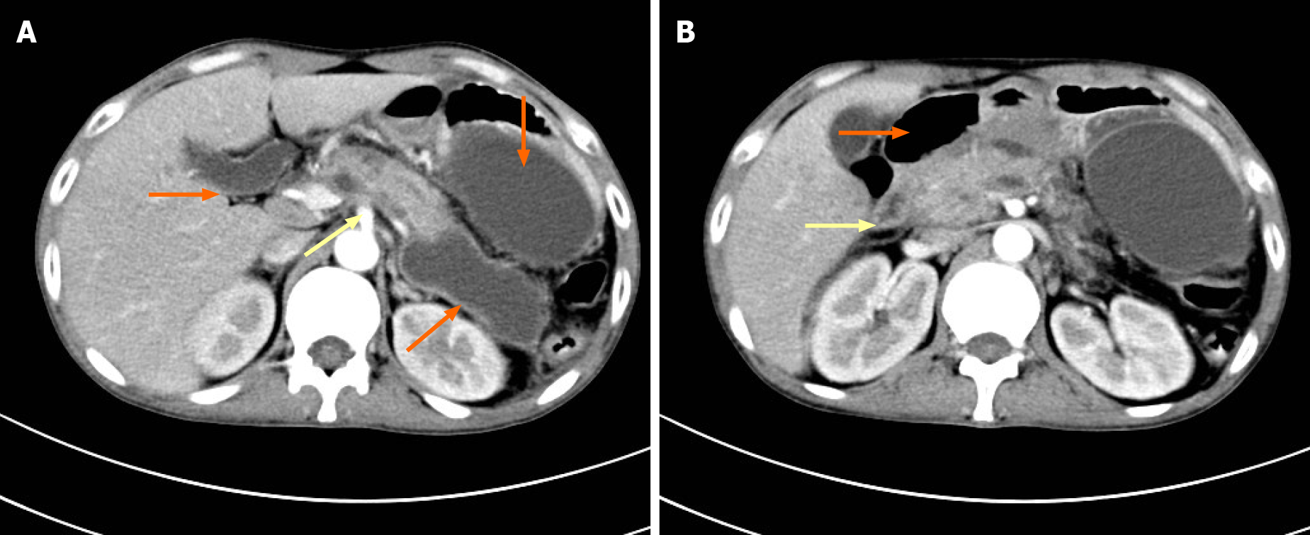
Figure 2 Computed tomography of the upper abdomen.
A: Computed tomography (CT) showing acute pancreatitis (yellow arrow) and multiple pancreatic pseudocysts (orange arrows); B: CT showing descendent duodenal edema (yellow arrow) and pneumatosis (orange arrow).
- Citation: Lu YL, Hu J, Zhang LY, Cen XY, Yang DH, Yu AY. Duodenal perforation after organophosphorus poisoning: A case report. World J Clin Cases 2021; 9(27): 8186-8191
- URL: https://www.wjgnet.com/2307-8960/full/v9/i27/8186.htm
- DOI: https://dx.doi.org/10.12998/wjcc.v9.i27.8186


