Copyright
©The Author(s) 2021.
World J Clin Cases. Sep 26, 2021; 9(27): 8186-8191
Published online Sep 26, 2021. doi: 10.12998/wjcc.v9.i27.8186
Published online Sep 26, 2021. doi: 10.12998/wjcc.v9.i27.8186
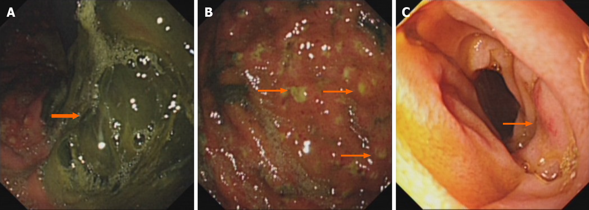
Figure 1 Gastroscopic images of the stomach and descending duodenum.
A: Gastroscopy showing that there are a large number of dark green secretions and membranous substances in the stomach cavity, which are mainly in the corner of the stomach and could not be washed and scraped off; B: Gastroscopy showing there are many bulges in the mucosa of the stomach cavity, purulent secretions at the top, and obvious congestion in the surrounding mucosa; C: Gastroscopy showing congestion and edema in part of the descending duodenum.
- Citation: Lu YL, Hu J, Zhang LY, Cen XY, Yang DH, Yu AY. Duodenal perforation after organophosphorus poisoning: A case report. World J Clin Cases 2021; 9(27): 8186-8191
- URL: https://www.wjgnet.com/2307-8960/full/v9/i27/8186.htm
- DOI: https://dx.doi.org/10.12998/wjcc.v9.i27.8186


