Copyright
©The Author(s) 2021.
World J Clin Cases. Sep 26, 2021; 9(27): 8071-8081
Published online Sep 26, 2021. doi: 10.12998/wjcc.v9.i27.8071
Published online Sep 26, 2021. doi: 10.12998/wjcc.v9.i27.8071
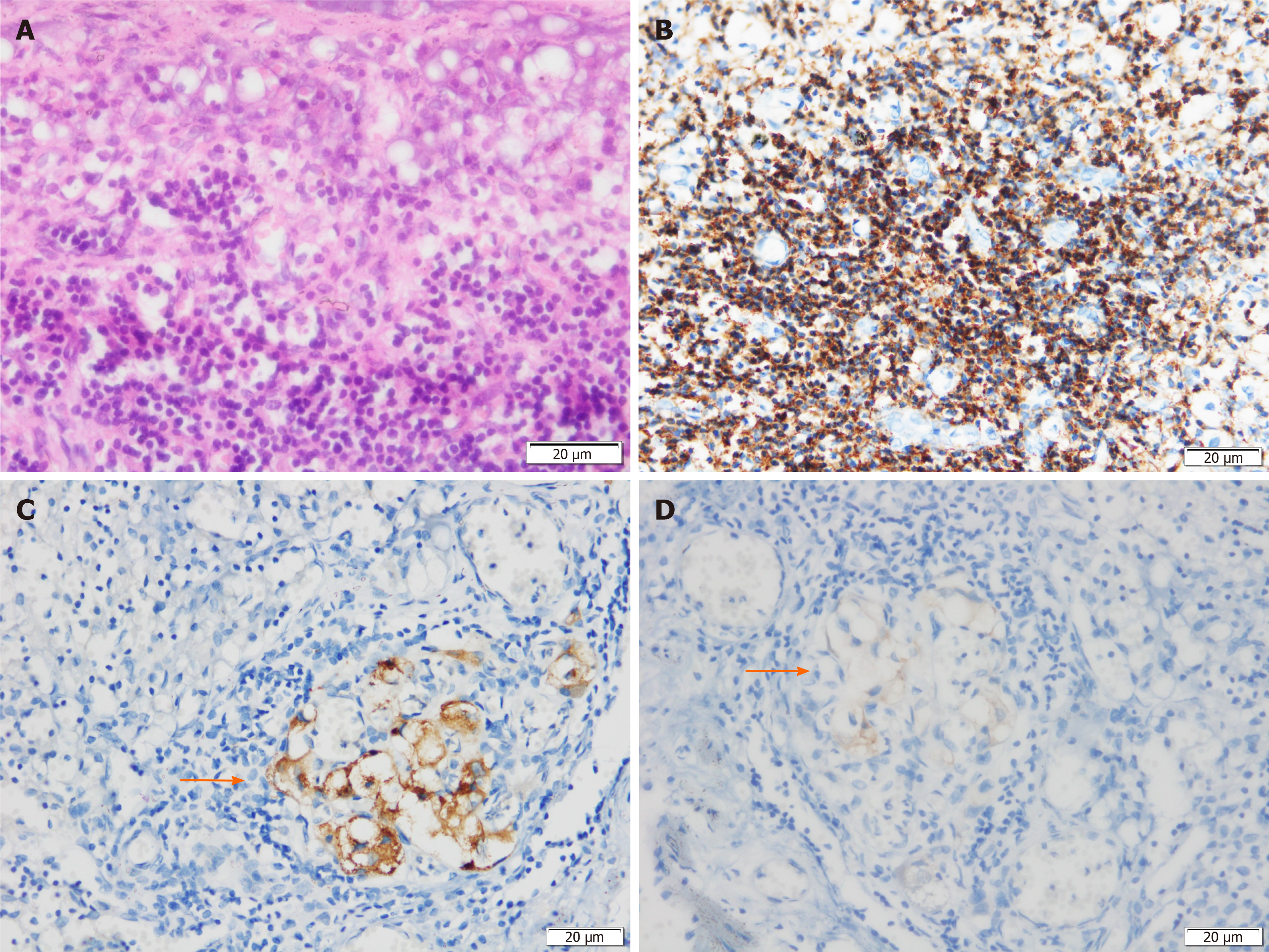
Figure 4 Histological and immunohistochemical examinations of the resected pancreatic draining lymph nodes.
A: Lymphatic tissue observation by hematoxylin-eosin staining; B: Positive staining for leukocyte common antigen confirms lymphatic tissue; C: The resected lymph nodes are positive for chromogranin A; D: The resected lymph nodes are positive for synaptophysin.
- Citation: Jiang CN, Cheng X, Shan J, Yang M, Xiao YQ. Primary pancreatic paraganglioma harboring lymph node metastasis: A case report. World J Clin Cases 2021; 9(27): 8071-8081
- URL: https://www.wjgnet.com/2307-8960/full/v9/i27/8071.htm
- DOI: https://dx.doi.org/10.12998/wjcc.v9.i27.8071


