Copyright
©The Author(s) 2021.
World J Clin Cases. Sep 26, 2021; 9(27): 8071-8081
Published online Sep 26, 2021. doi: 10.12998/wjcc.v9.i27.8071
Published online Sep 26, 2021. doi: 10.12998/wjcc.v9.i27.8071
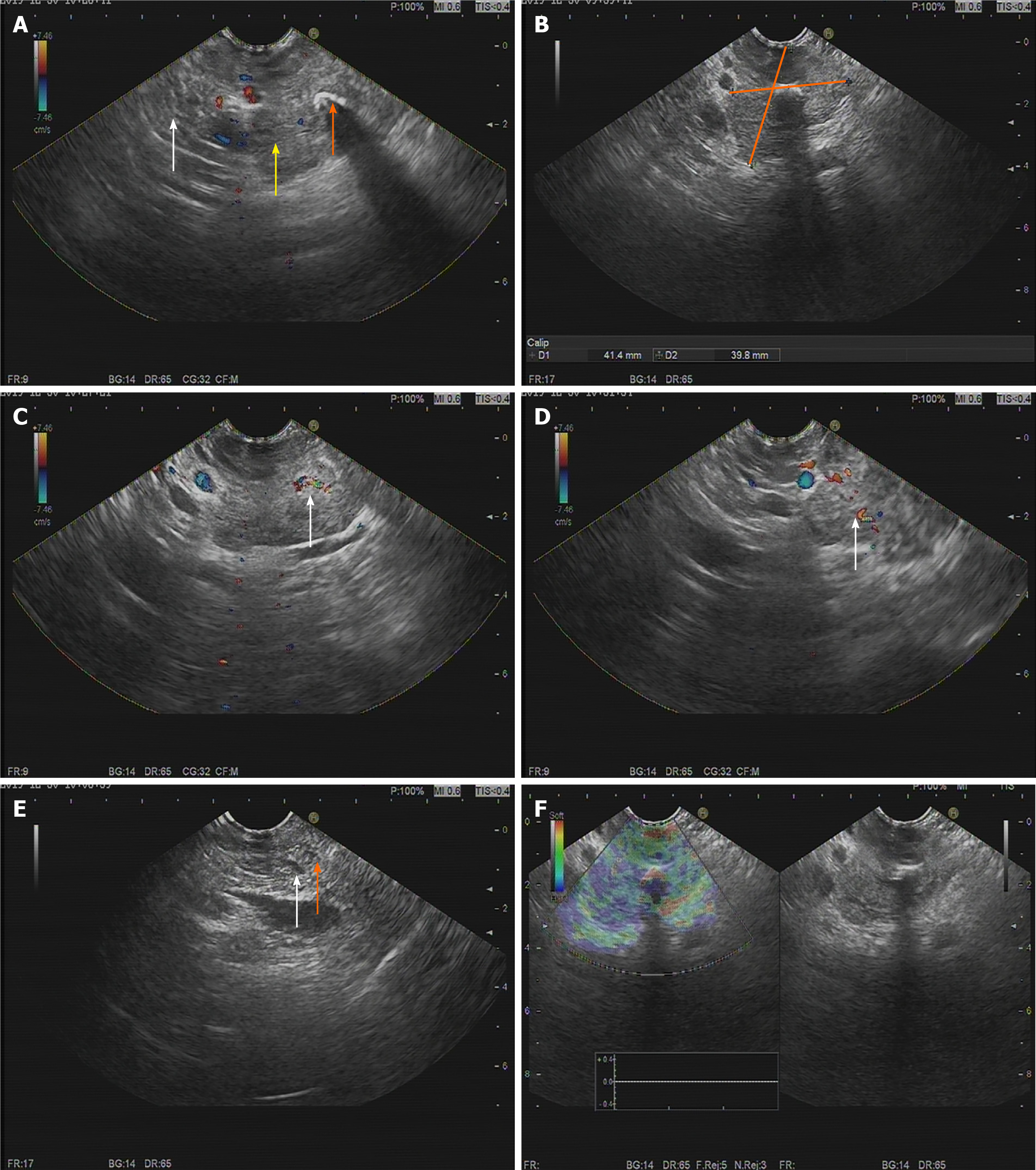
Figure 2 Endoscopic ultrasonography findings.
A: The tumor (yellow arrow) has an obscure boundary with the pancreas (white arrow), and nodular calcification can be seen inside the tumor (orange arrow); B: The tumor is 41.4 mm × 39.8 mm in size; C and D: A modest blood flow signal was detected inside the tumor (white arrow); E: No dilation of the bile duct (white arrow) and pancreatic duct (orange arrow); F: Elastography revealed the soft texture of the tumor.
- Citation: Jiang CN, Cheng X, Shan J, Yang M, Xiao YQ. Primary pancreatic paraganglioma harboring lymph node metastasis: A case report. World J Clin Cases 2021; 9(27): 8071-8081
- URL: https://www.wjgnet.com/2307-8960/full/v9/i27/8071.htm
- DOI: https://dx.doi.org/10.12998/wjcc.v9.i27.8071


