Copyright
©The Author(s) 2021.
World J Clin Cases. Sep 26, 2021; 9(27): 8071-8081
Published online Sep 26, 2021. doi: 10.12998/wjcc.v9.i27.8071
Published online Sep 26, 2021. doi: 10.12998/wjcc.v9.i27.8071
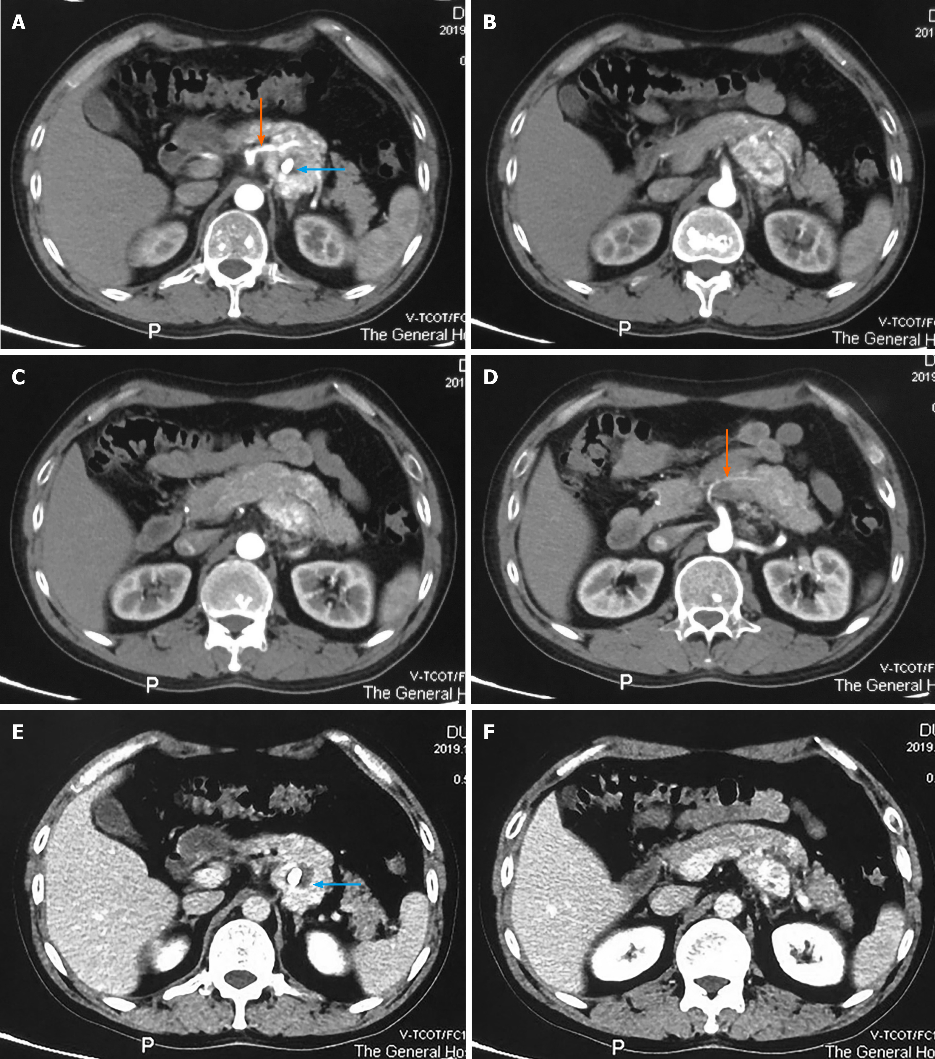
Figure 1 Abdominal contrast enhanced computed tomography findings.
A-D: The tumor shows marked enhancement during the arterial phase, the orange arrows show the vessels supplying the tumor from the superior mesenteric artery, and nodular calcification is observed inside the tumor (blue arrow); E and F: Enhancement is maintained, to a certain degree, during the venous phase, and nodular calcification was observed inside the tumor (blue arrow).
- Citation: Jiang CN, Cheng X, Shan J, Yang M, Xiao YQ. Primary pancreatic paraganglioma harboring lymph node metastasis: A case report. World J Clin Cases 2021; 9(27): 8071-8081
- URL: https://www.wjgnet.com/2307-8960/full/v9/i27/8071.htm
- DOI: https://dx.doi.org/10.12998/wjcc.v9.i27.8071


