Copyright
©The Author(s) 2021.
World J Clin Cases. Aug 6, 2021; 9(22): 6501-6509
Published online Aug 6, 2021. doi: 10.12998/wjcc.v9.i22.6501
Published online Aug 6, 2021. doi: 10.12998/wjcc.v9.i22.6501
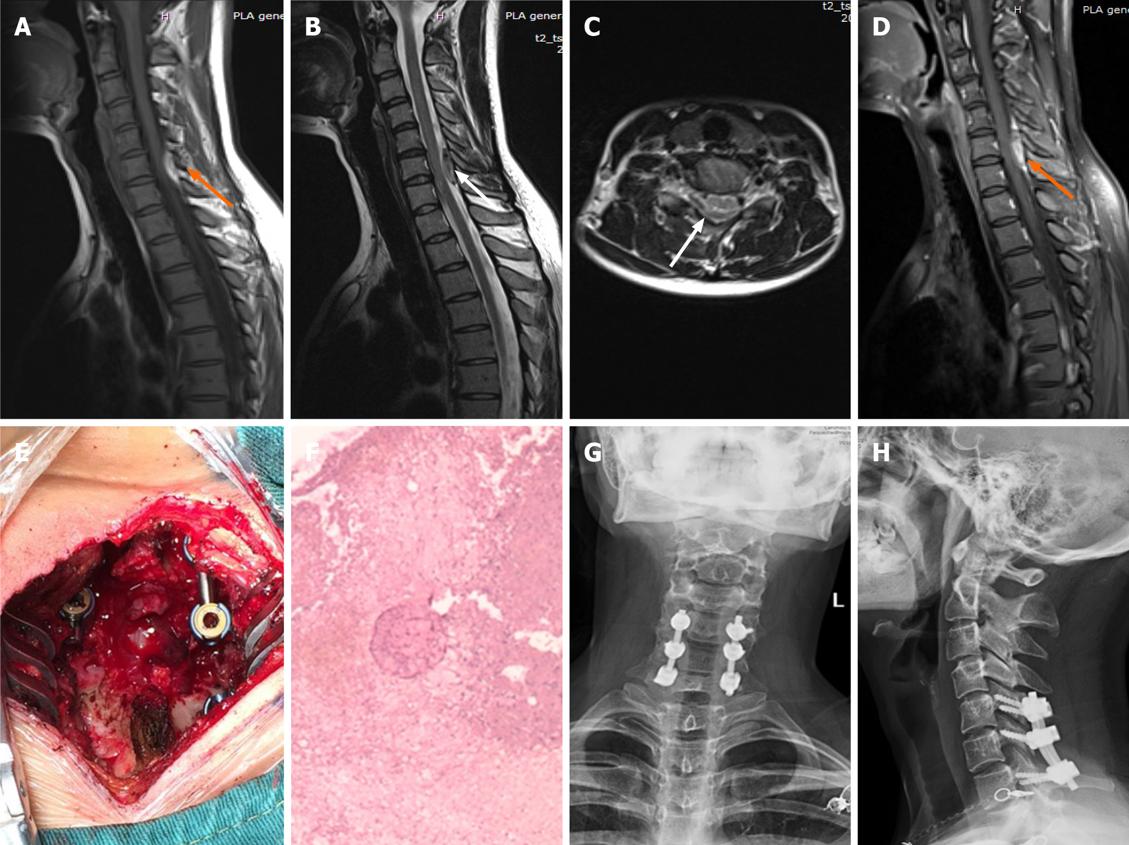
Figure 1 Imaging examinations of case 1.
A: T1-weighted preoperative magnetic resonance imaging (MRI) image shows high signal intensity (orange arrow); B and C: Preoperative T2-weighted image shows low signal intensity, and an axial T2-weighted image demonstrates that the hematoma occurred in the posterior region (white arrow); D: Preoperative enhanced MRI suggests an enhanced hematoma signal (orange arrow); E: Intraoperative photograph shows that spinal cord compression has recovered; F: Postoperative pathology suggested a hematoma; G and H: X-ray at the 3-mo follow-up indicated intact internal fixation.
- Citation: Liu H, Zhang T, Qu T, Yang CW, Li SK. Spinal epidural hematoma after spinal manipulation therapy: Report of three cases and a literature review. World J Clin Cases 2021; 9(22): 6501-6509
- URL: https://www.wjgnet.com/2307-8960/full/v9/i22/6501.htm
- DOI: https://dx.doi.org/10.12998/wjcc.v9.i22.6501


