Copyright
©The Author(s) 2021.
World J Clin Cases. Jul 26, 2021; 9(21): 6125-6129
Published online Jul 26, 2021. doi: 10.12998/wjcc.v9.i21.6125
Published online Jul 26, 2021. doi: 10.12998/wjcc.v9.i21.6125
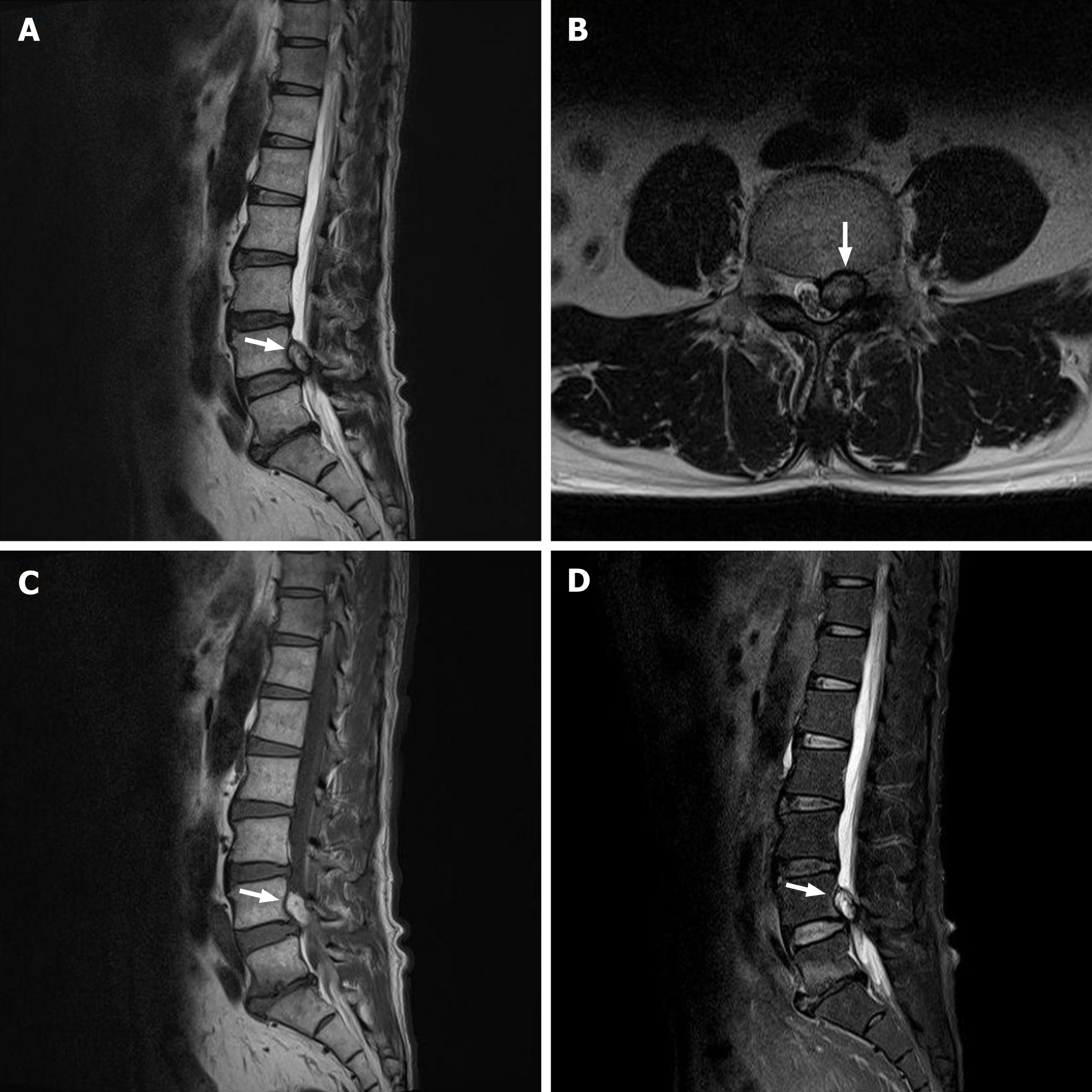
Figure 1 Magnetic resonance imaging reveals a mass lesion at the L4-5 level with thecal sac compression and encroachment of the left L4-5 neural foramen.
A and B: The T2-weighted images show mainly hyperintense with some heterogeneity. Also, T1 signal hyperintensity and lack of fat suppression were observed on the T1-weighted (C) and fat-suppressed T2-weigeted (D) images.
- Citation: Yu D, Lee W, Chang MC. Ligamentum flavum hematoma following a traffic accident: A case report. World J Clin Cases 2021; 9(21): 6125-6129
- URL: https://www.wjgnet.com/2307-8960/full/v9/i21/6125.htm
- DOI: https://dx.doi.org/10.12998/wjcc.v9.i21.6125


