Copyright
©The Author(s) 2021.
World J Clin Cases. Jul 26, 2021; 9(21): 6032-6040
Published online Jul 26, 2021. doi: 10.12998/wjcc.v9.i21.6032
Published online Jul 26, 2021. doi: 10.12998/wjcc.v9.i21.6032
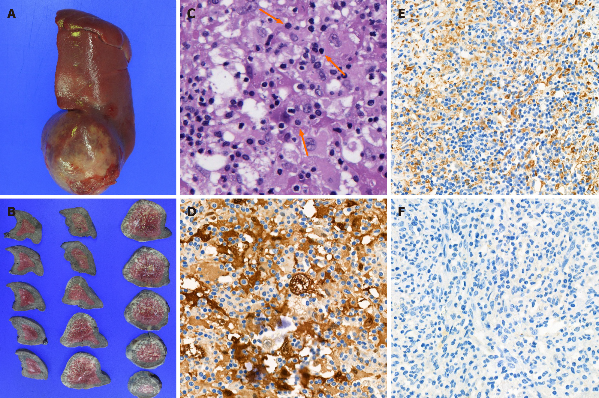
Figure 4 Pathologic images of splenic masses.
A: Resected specimen showed contour bulging splenic mass; B: Cut surface of the resected specimen; C: Hematoxylin and eosin-stained section (200 ×) showed histiocytic proliferation with emperipolesis (arrows); D: Proliferating histiocytes were positive for S-100 protein stained section (400 ×); E: Proliferating histiocytes were positive for CD68 antigen stained section (400 ×); F: proliferating histiocytes were negative for CD1a stained section (400 ×).
- Citation: Ryu H, Hwang JY, Kim YW, Kim TU, Jang JY, Park SE, Yang EJ, Shin DH. Rosai-Dorfman disease in the spleen of a pediatric patient: A case report. World J Clin Cases 2021; 9(21): 6032-6040
- URL: https://www.wjgnet.com/2307-8960/full/v9/i21/6032.htm
- DOI: https://dx.doi.org/10.12998/wjcc.v9.i21.6032


