Copyright
©The Author(s) 2021.
World J Clin Cases. Jul 26, 2021; 9(21): 6032-6040
Published online Jul 26, 2021. doi: 10.12998/wjcc.v9.i21.6032
Published online Jul 26, 2021. doi: 10.12998/wjcc.v9.i21.6032
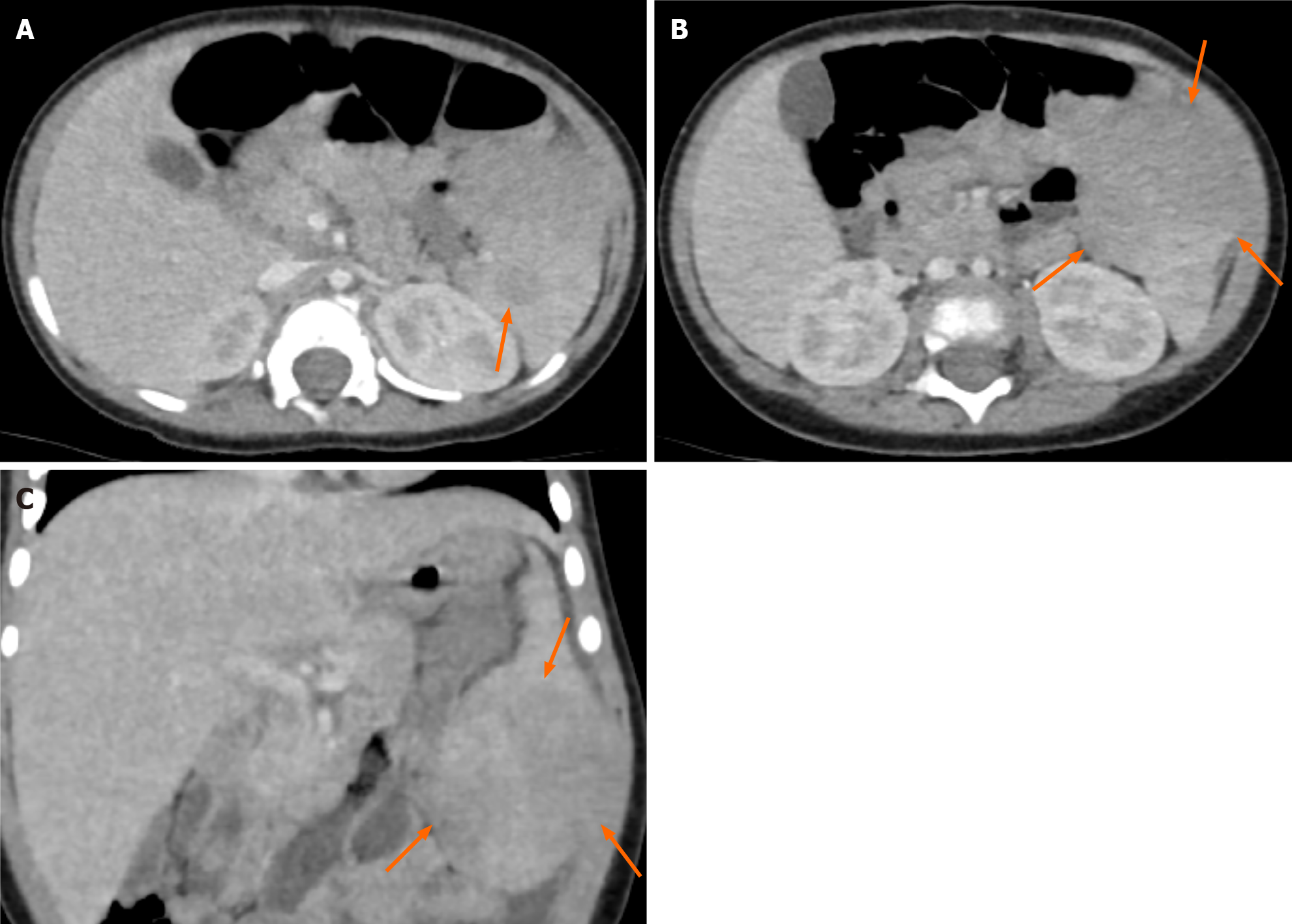
Figure 2 Computed tomography images of two solid splenic masses.
A: Axial image showed smaller hypodense mass; B: Axial image demonstrated larger exophytic hypodense mass; C: Coronal image revealed larger exophytic hypodense mass.
- Citation: Ryu H, Hwang JY, Kim YW, Kim TU, Jang JY, Park SE, Yang EJ, Shin DH. Rosai-Dorfman disease in the spleen of a pediatric patient: A case report. World J Clin Cases 2021; 9(21): 6032-6040
- URL: https://www.wjgnet.com/2307-8960/full/v9/i21/6032.htm
- DOI: https://dx.doi.org/10.12998/wjcc.v9.i21.6032


