Copyright
©The Author(s) 2021.
World J Clin Cases. Jun 16, 2021; 9(17): 4423-4432
Published online Jun 16, 2021. doi: 10.12998/wjcc.v9.i17.4423
Published online Jun 16, 2021. doi: 10.12998/wjcc.v9.i17.4423
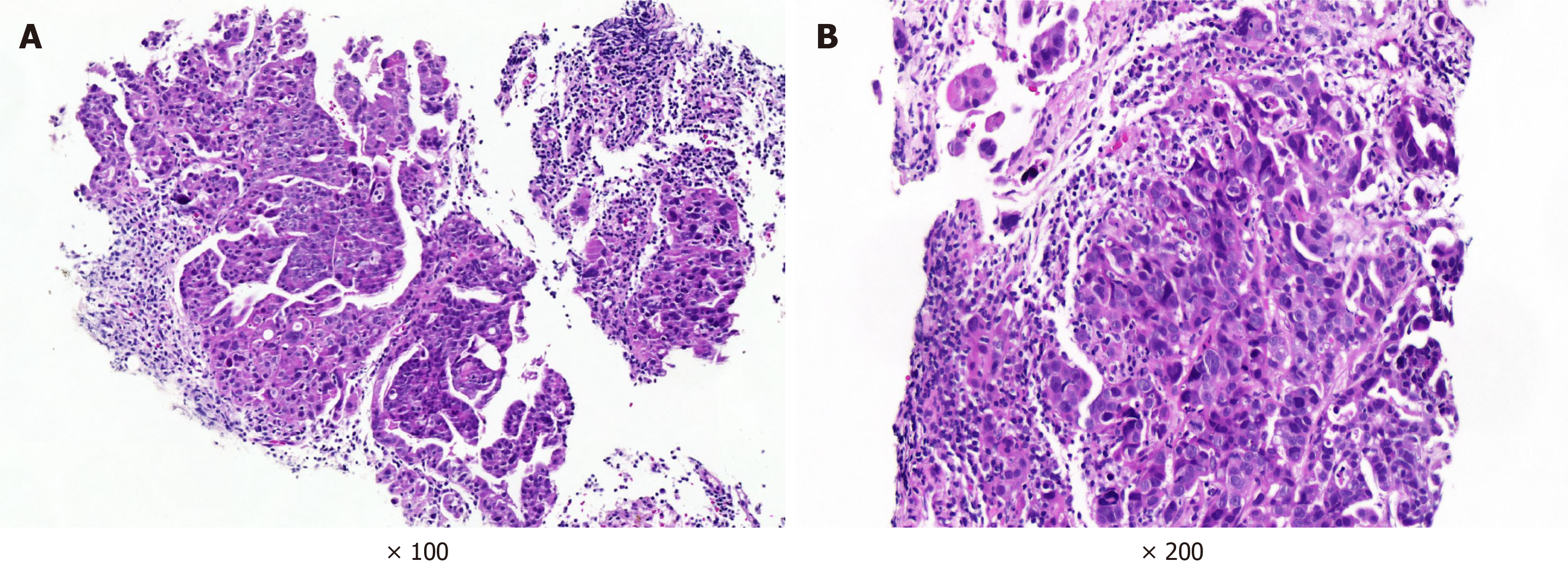
Figure 4 Lymph node pathology.
A: Left cervical lymph node pathology. Hematoxylin-eosin (HE) staining, × 100 magnification. The lymph nodes were infiltrated by atypical cells, and the atypical cells partly had an adenoid structure; B: Retroperitoneal lymph node pathology. HE staining, × 200 magnification. Atypical cells grew in nests, some of which had adenoid structures and infiltrating growth, with nuclear divisions visible
- Citation: Lou Y, Xu SH, Zhang SR, Shu QF, Liu XL. Anti-Yo antibody-positive paraneoplastic cerebellar degeneration in a patient with possible cholangiocarcinoma: A case report and review of the literature. World J Clin Cases 2021; 9(17): 4423-4432
- URL: https://www.wjgnet.com/2307-8960/full/v9/i17/4423.htm
- DOI: https://dx.doi.org/10.12998/wjcc.v9.i17.4423


