Copyright
©The Author(s) 2021.
World J Clin Cases. Jun 16, 2021; 9(17): 4423-4432
Published online Jun 16, 2021. doi: 10.12998/wjcc.v9.i17.4423
Published online Jun 16, 2021. doi: 10.12998/wjcc.v9.i17.4423
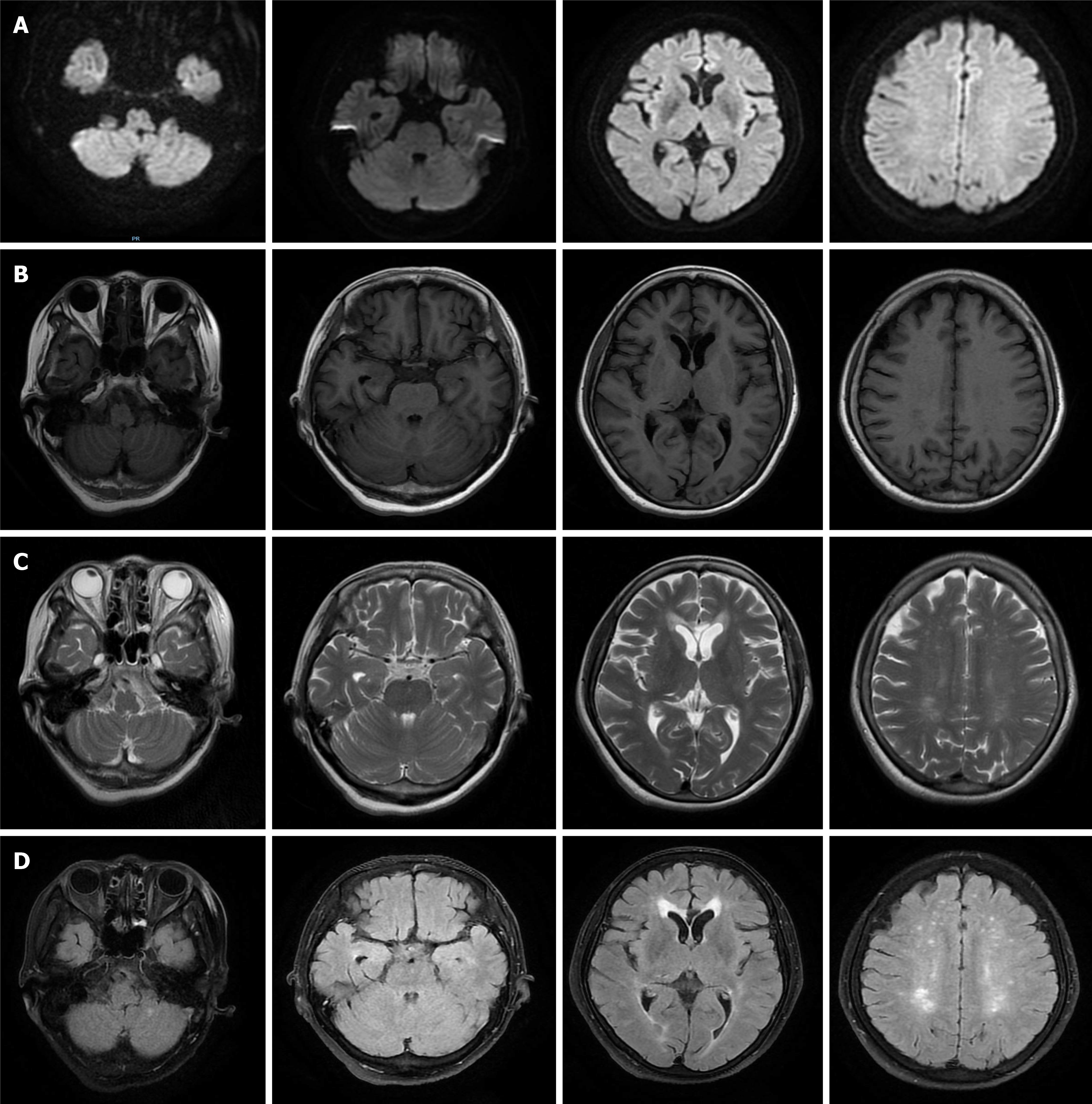
Figure 2 Magnetic resonance imaging showed multiple sub-frontal and parietal cortexes, semi-oval centers, and lateral ventricles with multiple spot-like and sheet-like lesions, equal signal on diffusion-weighted imaging, slightly longer signal on T1, longer signal on T2, and partially high signal on flair.
A: Diffusion-weighted imaging; B: T1; C: T2; D: Flair.
- Citation: Lou Y, Xu SH, Zhang SR, Shu QF, Liu XL. Anti-Yo antibody-positive paraneoplastic cerebellar degeneration in a patient with possible cholangiocarcinoma: A case report and review of the literature. World J Clin Cases 2021; 9(17): 4423-4432
- URL: https://www.wjgnet.com/2307-8960/full/v9/i17/4423.htm
- DOI: https://dx.doi.org/10.12998/wjcc.v9.i17.4423


