Copyright
©The Author(s) 2021.
World J Clin Cases. Jun 16, 2021; 9(17): 4348-4356
Published online Jun 16, 2021. doi: 10.12998/wjcc.v9.i17.4348
Published online Jun 16, 2021. doi: 10.12998/wjcc.v9.i17.4348
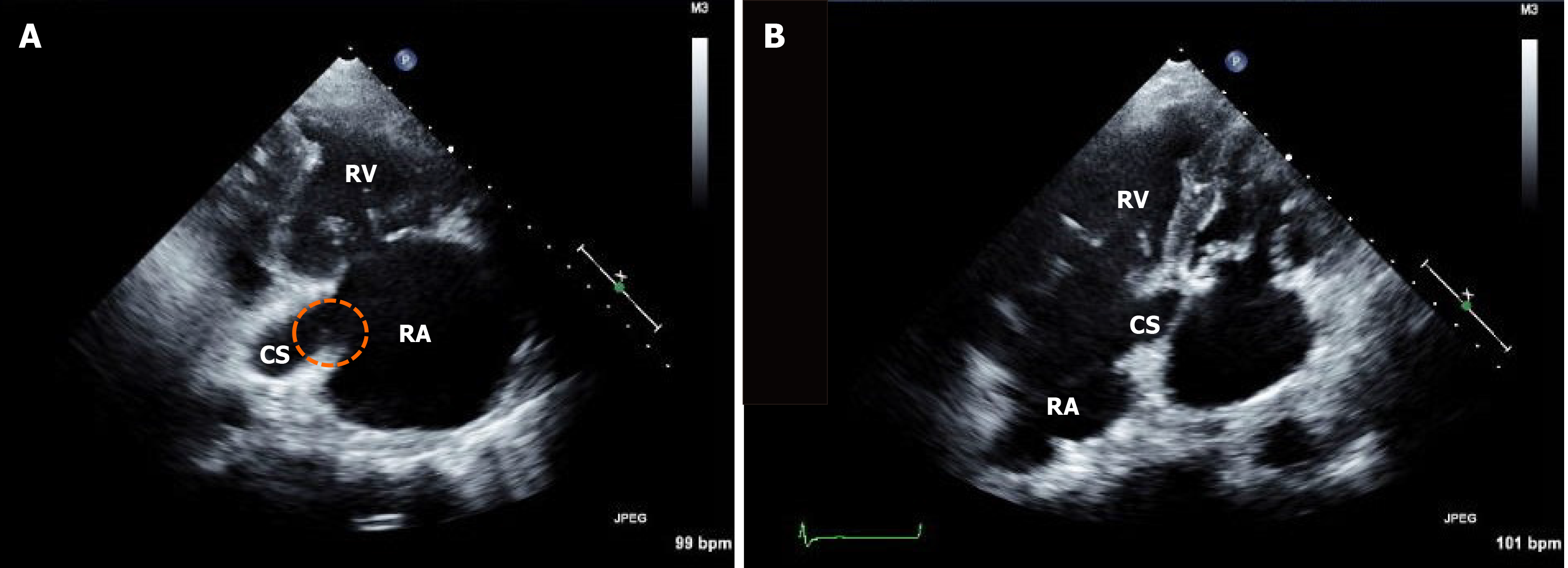
Figure 3 Follow-up echocardiographic imaging.
A: Right ventricular inflow; B: Modified apical four-chamber views showing a remnant vegetation at the coronary sinus ostium (dotted circle). RA: Right atrium; RV: Right ventricle; CS: Coronary sinus.
- Citation: Hwang HJ, Kang SW. Coronary sinus endocarditis in a hemodialysis patient: A case report and review of literature. World J Clin Cases 2021; 9(17): 4348-4356
- URL: https://www.wjgnet.com/2307-8960/full/v9/i17/4348.htm
- DOI: https://dx.doi.org/10.12998/wjcc.v9.i17.4348


