Copyright
©The Author(s) 2021.
World J Clin Cases. Jun 6, 2021; 9(16): 3979-3987
Published online Jun 6, 2021. doi: 10.12998/wjcc.v9.i16.3979
Published online Jun 6, 2021. doi: 10.12998/wjcc.v9.i16.3979
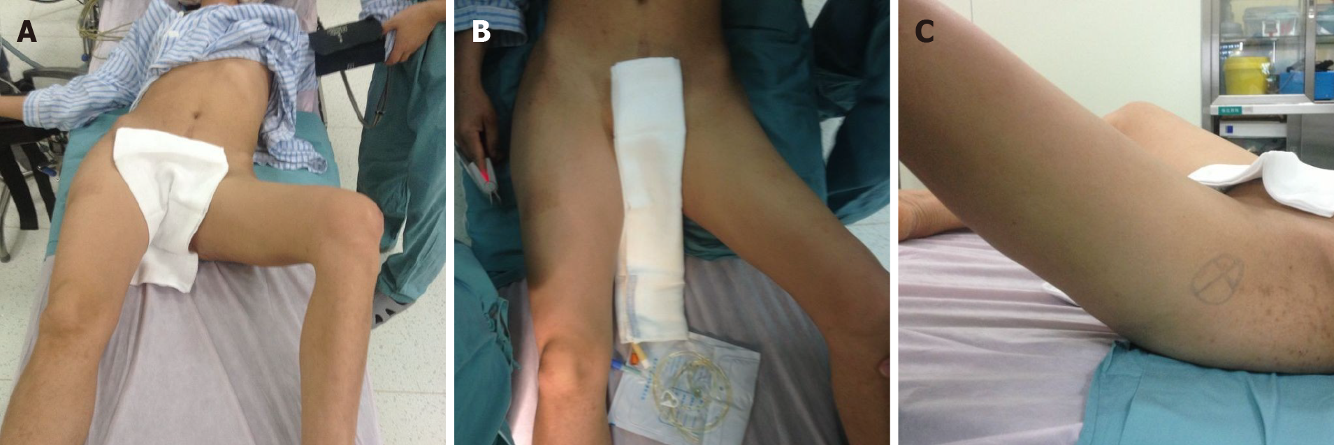
Figure 1 Physical examination.
A: Front view of the patient showing that there was compensatory scoliosis towards the left, and the pelvis was lower on the left side; B and C: Front and lateral view of the left hip showing that the hip was fixed in 40° of flexion, 45° of abduction, and 30° of external rotation.
- Citation: Li WZ, Wang JJ, Ni JD, Song DY, Ding ML, Huang J, He GX. Old unreduced obturator dislocation of the hip: A case report. World J Clin Cases 2021; 9(16): 3979-3987
- URL: https://www.wjgnet.com/2307-8960/full/v9/i16/3979.htm
- DOI: https://dx.doi.org/10.12998/wjcc.v9.i16.3979


