Copyright
©The Author(s) 2021.
World J Clin Cases. May 16, 2021; 9(14): 3350-3355
Published online May 16, 2021. doi: 10.12998/wjcc.v9.i14.3350
Published online May 16, 2021. doi: 10.12998/wjcc.v9.i14.3350
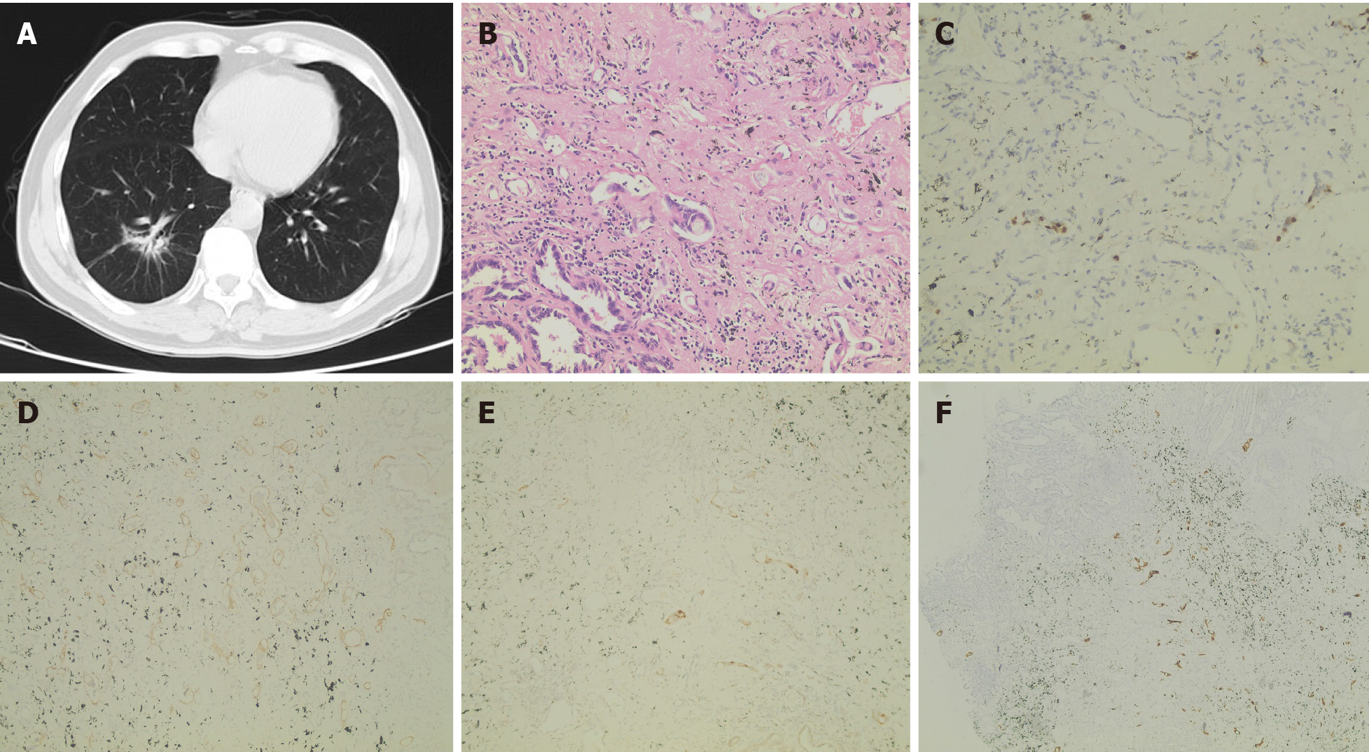
Figure 1 Patient disease diagnosis.
A: Computed tomography image of lung tissue; B: Typical image of postoperative pathology; C-F: Immunohistochemical staining images of Ki67 (+) (C), D2-40 (+) (D), MUC5AC (+) (E), and Villin (+) (F).
- Citation: Wu ZW, Sha Y, Chen Q, Hou J, Sun Y, Lu WK, Chen J, Yu LJ. Novel intergenic KIF5B-MET fusion variant in a patient with gastric cancer: A case report. World J Clin Cases 2021; 9(14): 3350-3355
- URL: https://www.wjgnet.com/2307-8960/full/v9/i14/3350.htm
- DOI: https://dx.doi.org/10.12998/wjcc.v9.i14.3350


