Copyright
©The Author(s) 2021.
World J Clin Cases. Apr 16, 2021; 9(11): 2611-2618
Published online Apr 16, 2021. doi: 10.12998/wjcc.v9.i11.2611
Published online Apr 16, 2021. doi: 10.12998/wjcc.v9.i11.2611
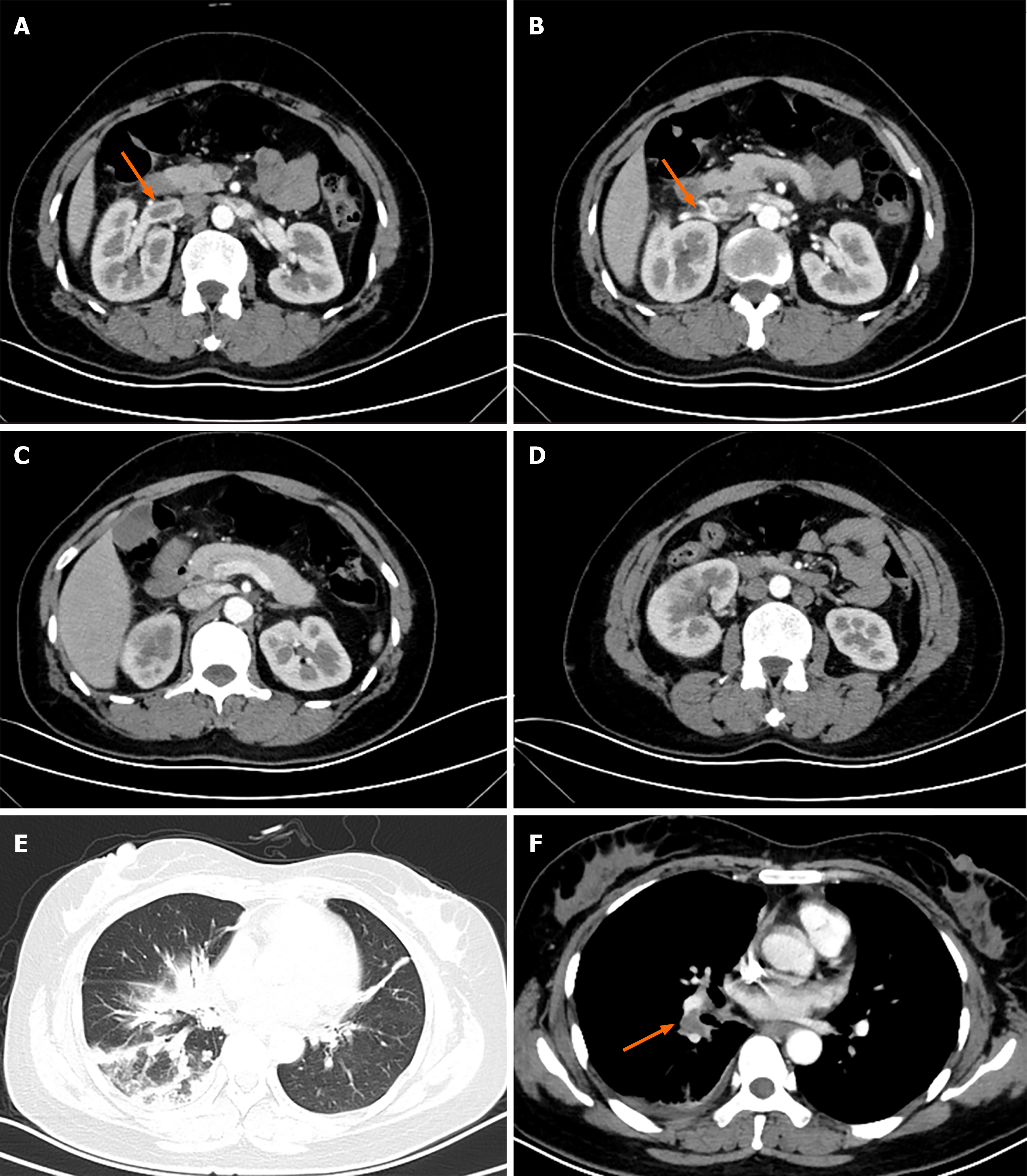
Figure 1 Computed tomography studies of the abdomen and chest on admission, with enhancement (intravenous contrast).
A and B: Abdominal computed tomography (CT) demonstrating a near-occlusive thrombus of the right renal vein (arrow) extending to the inferior vena cava; C and D: Enlargement of the right kidney on abdominal CT, with upper and lower poles showing poor perfusion; E: Chest CT with patchy infiltrates in lung window and right pleural effusion; F: Emboli of the right pulmonary artery (orange arrow) on chest CT.
- Citation: Wu C, Zhou XM, Liu XD. Eltrombopag-related renal vein thromboembolism in a patient with immune thrombocytopenia: A case report. World J Clin Cases 2021; 9(11): 2611-2618
- URL: https://www.wjgnet.com/2307-8960/full/v9/i11/2611.htm
- DOI: https://dx.doi.org/10.12998/wjcc.v9.i11.2611


