Copyright
©The Author(s) 2021.
World J Clin Cases. Apr 6, 2021; 9(10): 2344-2351
Published online Apr 6, 2021. doi: 10.12998/wjcc.v9.i10.2344
Published online Apr 6, 2021. doi: 10.12998/wjcc.v9.i10.2344
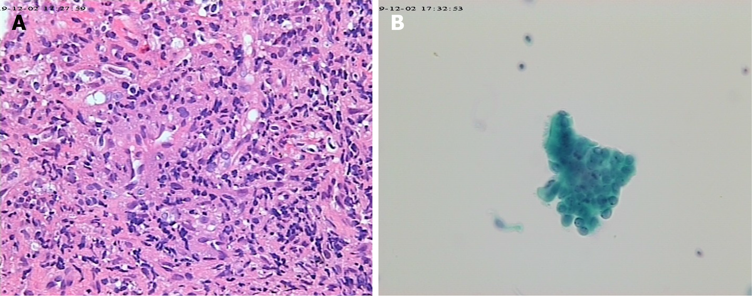
Figure 3 Results of bronchoscopic biopsy.
A: Biopsies revealed local granulomatous structure; peripheral fibrous hyperplasia; lymphocyte, plasma cell and neutrophil infiltration (Papanicolaou stain, 20 ×); B: By microscopy, columnar epithelial cells and individual lymphocytes were seen, and no heterotypic cells were found (hematoxylin and eosin stain, 20 ×).
- Citation: Li XJ, Yang L, Yan XF, Zhan CT, Liu JH. Granulomatosis with polyangiitis presenting as high fever with diffuse alveolar hemorrhage and otitis media: A case report. World J Clin Cases 2021; 9(10): 2344-2351
- URL: https://www.wjgnet.com/2307-8960/full/v9/i10/2344.htm
- DOI: https://dx.doi.org/10.12998/wjcc.v9.i10.2344


