Copyright
©The Author(s) 2020.
World J Clin Cases. Mar 26, 2020; 8(6): 1150-1157
Published online Mar 26, 2020. doi: 10.12998/wjcc.v8.i6.1150
Published online Mar 26, 2020. doi: 10.12998/wjcc.v8.i6.1150
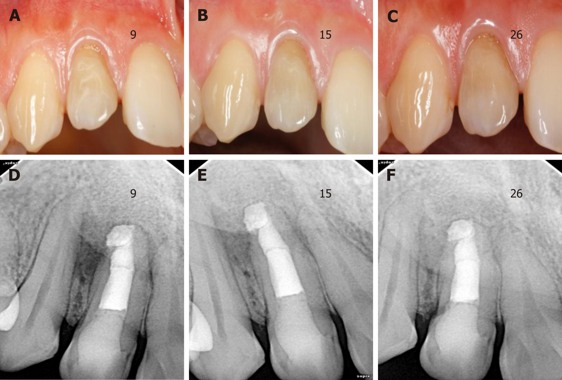
Figure 4 Follow-up images at different periods.
A-C: The labial view of the invaginated tooth during 9-, 15- and 26-mo follow-up examinations; D-F: Periapical radiographs taken at the 9-, 15- and 26-mo follow-up examinations. The apical radiolucency was significantly reduced.
- Citation: Liu J, Zhang YR, Zhang FY, Zhang GD, Xu H. Microscopic removal of type III dens invaginatus and preparation of apical barrier with mineral trioxide aggregate in a maxillary lateral incisor: A case report and review of literature. World J Clin Cases 2020; 8(6): 1150-1157
- URL: https://www.wjgnet.com/2307-8960/full/v8/i6/1150.htm
- DOI: https://dx.doi.org/10.12998/wjcc.v8.i6.1150


