Copyright
©The Author(s) 2020.
World J Clin Cases. Mar 26, 2020; 8(6): 1150-1157
Published online Mar 26, 2020. doi: 10.12998/wjcc.v8.i6.1150
Published online Mar 26, 2020. doi: 10.12998/wjcc.v8.i6.1150
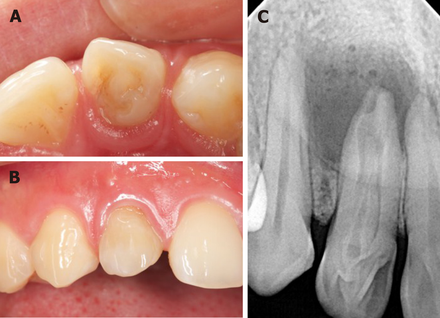
Figure 1 The intraoral examination before treatment.
A: The lingual view: the tooth has unusual morphologic features; B: The labial view: the upper right lateral tooth is slightly discolored; C: A preoperative radiograph of the tooth showed a type III dens invaginatus and an associated large apical radiolucency.
- Citation: Liu J, Zhang YR, Zhang FY, Zhang GD, Xu H. Microscopic removal of type III dens invaginatus and preparation of apical barrier with mineral trioxide aggregate in a maxillary lateral incisor: A case report and review of literature. World J Clin Cases 2020; 8(6): 1150-1157
- URL: https://www.wjgnet.com/2307-8960/full/v8/i6/1150.htm
- DOI: https://dx.doi.org/10.12998/wjcc.v8.i6.1150


