Copyright
©The Author(s) 2020.
World J Clin Cases. Mar 6, 2020; 8(5): 932-938
Published online Mar 6, 2020. doi: 10.12998/wjcc.v8.i5.932
Published online Mar 6, 2020. doi: 10.12998/wjcc.v8.i5.932
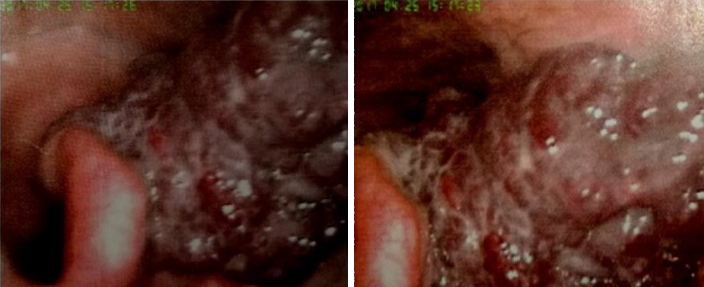
Figure 1 Preoperative laryngoscopic examination showing a hypopharyngeal hemangioma.
A submucosal vascular lesion extending into the epiglottis, left arytenoid cartilage, lateral to the aryepiglottic fold, and pyriform sinus was visualized.
- Citation: Jin M, Wang CY, Da YX, Zhu W, Jiang H. Surgical resection of a large hypopharyngeal hemangioma in an adult using neodymium-doped yttrium aluminum garnet laser: A case report. World J Clin Cases 2020; 8(5): 932-938
- URL: https://www.wjgnet.com/2307-8960/full/v8/i5/932.htm
- DOI: https://dx.doi.org/10.12998/wjcc.v8.i5.932


