Copyright
©The Author(s) 2020.
World J Clin Cases. Feb 26, 2020; 8(4): 806-814
Published online Feb 26, 2020. doi: 10.12998/wjcc.v8.i4.806
Published online Feb 26, 2020. doi: 10.12998/wjcc.v8.i4.806
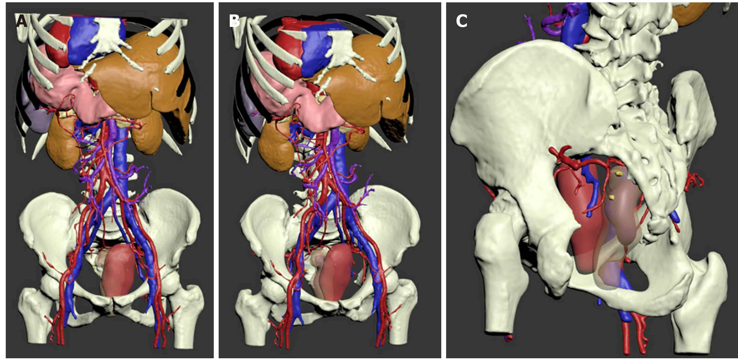
Figure 6 The three-dimensional model of abdominal organs and blood vessels of the patient.
A: Positive view. The patient's heart, liver, stomach, pancreas, spleen, and large blood vessels were visible as "mirror human" images; B: Starboard forward view. It demonstrates rectum and its surrounding organs, blood vessels, and lymph nodes; C: Left pelvic elevation view. The anterior is the uterus in pink, and the posterior translucent material is the rectum. The darker lump inside the rectum lumen shows the tumor. The yellow nodules around the rectum are metastatic lymph nodes.
- Citation: Chen T, Que YT, Zhang YH, Long FY, Li Y, Huang X, Wang YN, Hu YF, Yu J, Li GX. Using Materialise's interactive medical image control system to reconstruct a model of a patient with rectal cancer and situs inversus totalis: A case report. World J Clin Cases 2020; 8(4): 806-814
- URL: https://www.wjgnet.com/2307-8960/full/v8/i4/806.htm
- DOI: https://dx.doi.org/10.12998/wjcc.v8.i4.806


