Copyright
©The Author(s) 2020.
World J Clin Cases. Dec 26, 2020; 8(24): 6480-6486
Published online Dec 26, 2020. doi: 10.12998/wjcc.v8.i24.6480
Published online Dec 26, 2020. doi: 10.12998/wjcc.v8.i24.6480
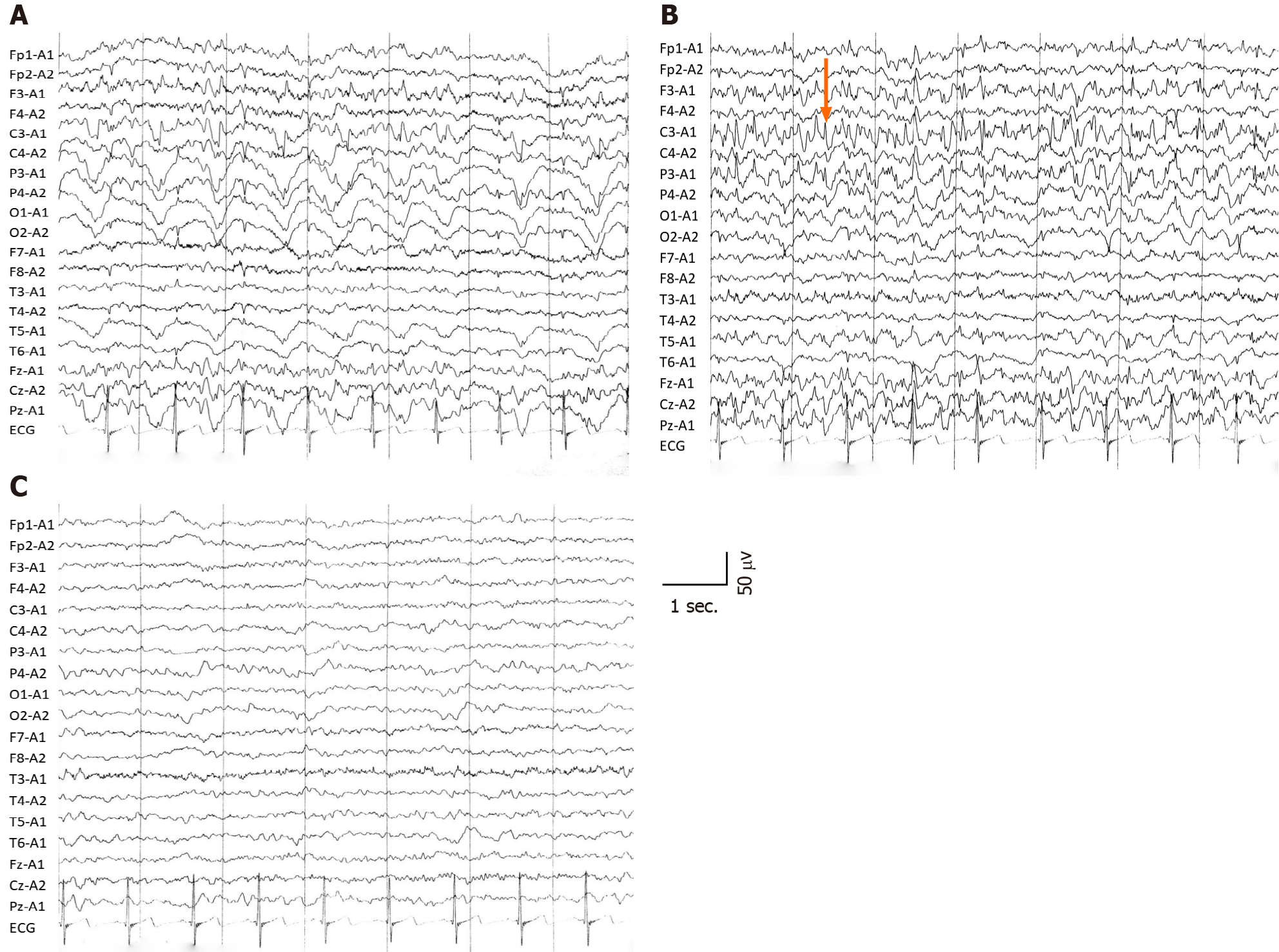
Figure 2 Electroencephalography showing different wave rhythms.
A: Interictal electroencephalography showed slow wave rhythm activity; B: Epileptiform discharge (orange arrow) in left parietooccipital during ictal period, which corresponded well to the clinical symptoms; C: The slow waves rhythm and background activity improved after 2 mo.
- Citation: Cui B, Wei L, Sun LY, Qu W, Zeng ZG, Liu Y, Zhu ZJ. Status epilepticus as an initial manifestation of hepatic encephalopathy: A case report. World J Clin Cases 2020; 8(24): 6480-6486
- URL: https://www.wjgnet.com/2307-8960/full/v8/i24/6480.htm
- DOI: https://dx.doi.org/10.12998/wjcc.v8.i24.6480


