Copyright
©The Author(s) 2020.
World J Clin Cases. Dec 6, 2020; 8(23): 6095-6102
Published online Dec 6, 2020. doi: 10.12998/wjcc.v8.i23.6095
Published online Dec 6, 2020. doi: 10.12998/wjcc.v8.i23.6095
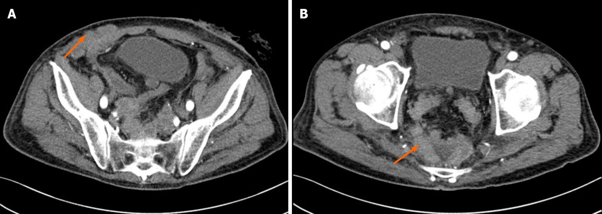
Figure 5 Contrast-enhanced computed tomography imaging scans 1 mo after surgery.
A: Computed tomography imaging showed that there were multiple nodules in the lower abdomen, which was consistent with our physical examination; B: There were many new lesions in the pelvis (orange arrow) compared with Figure 2B.
- Citation: Chen ZZ, Huang W, Wei ZQ. Small-cell neuroendocrine carcinoma of the rectum — a rare tumor type with poor prognosis: A case report and review of literature. World J Clin Cases 2020; 8(23): 6095-6102
- URL: https://www.wjgnet.com/2307-8960/full/v8/i23/6095.htm
- DOI: https://dx.doi.org/10.12998/wjcc.v8.i23.6095


