Copyright
©The Author(s) 2020.
World J Clin Cases. Nov 26, 2020; 8(22): 5555-5563
Published online Nov 26, 2020. doi: 10.12998/wjcc.v8.i22.5555
Published online Nov 26, 2020. doi: 10.12998/wjcc.v8.i22.5555
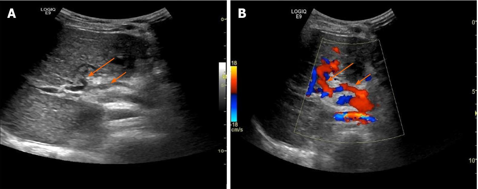
Figure 4 Grayscale and color Doppler ultrasonography of an 8-year-old girl after recanalized umbilical vein as a conduit for Rex shunt.
A: Grayscale ultrasonography; B: Color Doppler ultrasonography. The girl was admitted to hospital with intermittent hematemesis and black stool. Bypassing the main portal vein with the umbilical vein was conducted through the splenic vein. The bypass vessel (splenic vein) and anastomoses were clearly displayed 7 d after operation. Long arrow indicates umbilical vein and short arrow indicates splenic vein.
- Citation: Zhang YQ, Wang Q, Wu M, Li Y, Wei XL, Zhang FX, Li Y, Shao GR, Xiao J. Sonographic features of umbilical vein recanalization for a Rex shunt on cavernous transformation of portal vein in children . World J Clin Cases 2020; 8(22): 5555-5563
- URL: https://www.wjgnet.com/2307-8960/full/v8/i22/5555.htm
- DOI: https://dx.doi.org/10.12998/wjcc.v8.i22.5555


