Copyright
©The Author(s) 2020.
World J Clin Cases. Nov 6, 2020; 8(21): 5149-5158
Published online Nov 6, 2020. doi: 10.12998/wjcc.v8.i21.5149
Published online Nov 6, 2020. doi: 10.12998/wjcc.v8.i21.5149
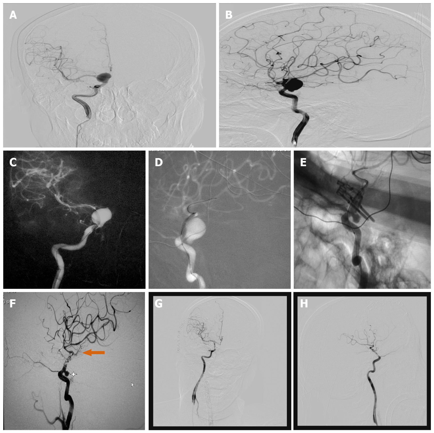
Figure 2 Preoperative, intraoperative and postoperative imaging of a 54-year-old female patient with a large carotid-ophthalmic aneurysm.
A and B: Pre-operative digital subtraction angiography (DSA) in anteroposterior (AP) and lateral (LAT) views: the giant stained aneurysm in the internal carotid-ophthalmic segment was visible, maximal diameter (20.4 mm), neck size (13.4 mm) and dome-neck ratio of 1.6; C and D: First intraoperative DSA (AP and LAT view) showed that the balloon was placed in the aneurysm neck; E and F: Second intraoperative DSA showed that the bulk of the aneurysm was clipped and not visualized, but a small portion of residual aneurysm was visualized; G and H: After adjusting the clips, third intraoperative DSA displayed that the aneurysm was completely clipped, but the parent artery was slightly narrowed due to the severe calcification of the aneurysmal neck.
- Citation: Zhang N, Xin WQ. Application of hybrid operating rooms for clipping large or giant intracranial carotid-ophthalmic aneurysms. World J Clin Cases 2020; 8(21): 5149-5158
- URL: https://www.wjgnet.com/2307-8960/full/v8/i21/5149.htm
- DOI: https://dx.doi.org/10.12998/wjcc.v8.i21.5149


