Copyright
©The Author(s) 2020.
World J Clin Cases. Oct 26, 2020; 8(20): 4895-4901
Published online Oct 26, 2020. doi: 10.12998/wjcc.v8.i20.4895
Published online Oct 26, 2020. doi: 10.12998/wjcc.v8.i20.4895
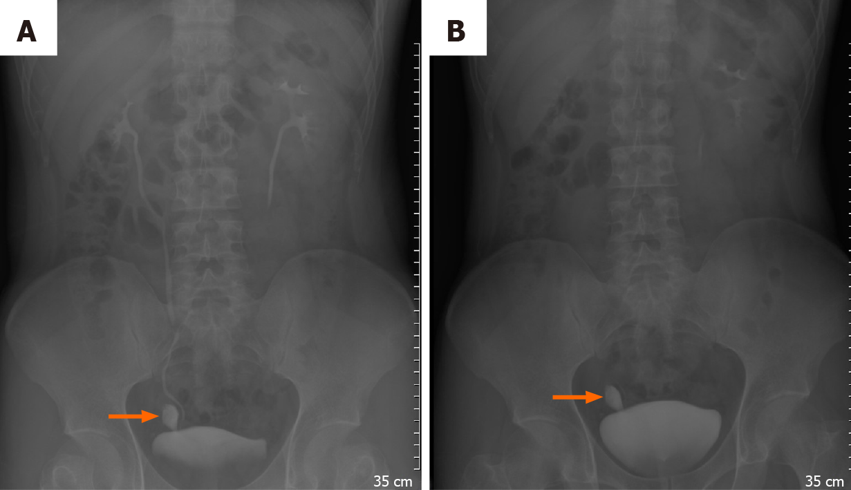
Figure 1 Intravenous pyelography was further investigated, and on 5-min film a well-defined contrast shape structure was observed; at 30-min film, the contrast was still trapped insides.
Differential diagnosis could be bladder rupture, ureteral tear, or anatomical abnormality. A: 5-min film; and B: 30-min film.
- Citation: Yang CH, Lin YS, Ou YC, Weng WC, Huang LH, Lu CH, Hsu CY, Tung MC. Adult metaplastic hutch diverticulum with robotic-assisted diverticulectomy and reconstruction: A case report. World J Clin Cases 2020; 8(20): 4895-4901
- URL: https://www.wjgnet.com/2307-8960/full/v8/i20/4895.htm
- DOI: https://dx.doi.org/10.12998/wjcc.v8.i20.4895


