
















©The Author(s) 2020.
World J Clin Cases. Sep 26, 2020; 8(18): 4114-4121
Published online Sep 26, 2020. doi: 10.12998/wjcc.v8.i18.4114
Published online Sep 26, 2020. doi: 10.12998/wjcc.v8.i18.4114
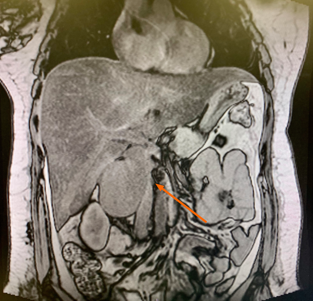
Figure 1 Magnetic resonance imaging of the coronal section showing a large tumor mass compressing the lateral wall of the inferior vena cava.
- Citation: Ribeiro Jr MAF, Elias YGB, Augusto SS, Néder PR, Costa CTK, Maurício AD, Sampaio AP, Fonseca AZ. Laparoscopic resection of primary retroperitoneal schwannoma: A case report. World J Clin Cases 2020; 8(18): 4114-4121
- URL: https://www.wjgnet.com/2307-8960/full/v8/i18/4114.htm
- DOI: https://dx.doi.org/10.12998/wjcc.v8.i18.4114