Copyright
©The Author(s) 2019.
World J Clin Cases. Feb 6, 2019; 7(3): 347-356
Published online Feb 6, 2019. doi: 10.12998/wjcc.v7.i3.347
Published online Feb 6, 2019. doi: 10.12998/wjcc.v7.i3.347
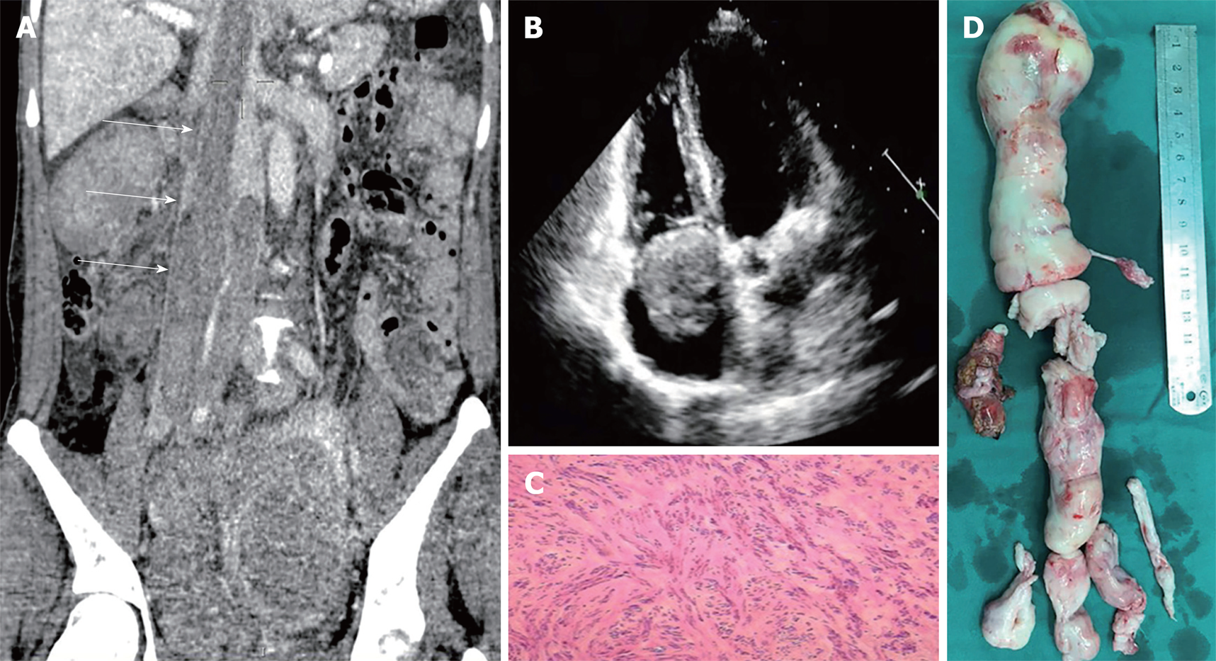
Figure 3 Representative images of Case 3.
A: Computed tomography scan shows a continuous mass arising from the pelvis through the inferior cava vein; B: Echocardiography shows the mass in the right cardiac chamber; C: Pathological examination of the tumor showed concentration of smooth muscle cells with local hyaline degeneration; D: The resected tumor (displayed from the top to the bottom).
- Citation: He J, Chen ZB, Wang SM, Liu MB, Li ZG, Li HY, Zhao G. Intravenous leiomyomatosis with different surgical approaches: Three case reports. World J Clin Cases 2019; 7(3): 347-356
- URL: https://www.wjgnet.com/2307-8960/full/v7/i3/347.htm
- DOI: https://dx.doi.org/10.12998/wjcc.v7.i3.347


