Copyright
©The Author(s) 2019.
World J Clin Cases. Nov 26, 2019; 7(22): 3872-3880
Published online Nov 26, 2019. doi: 10.12998/wjcc.v7.i22.3872
Published online Nov 26, 2019. doi: 10.12998/wjcc.v7.i22.3872
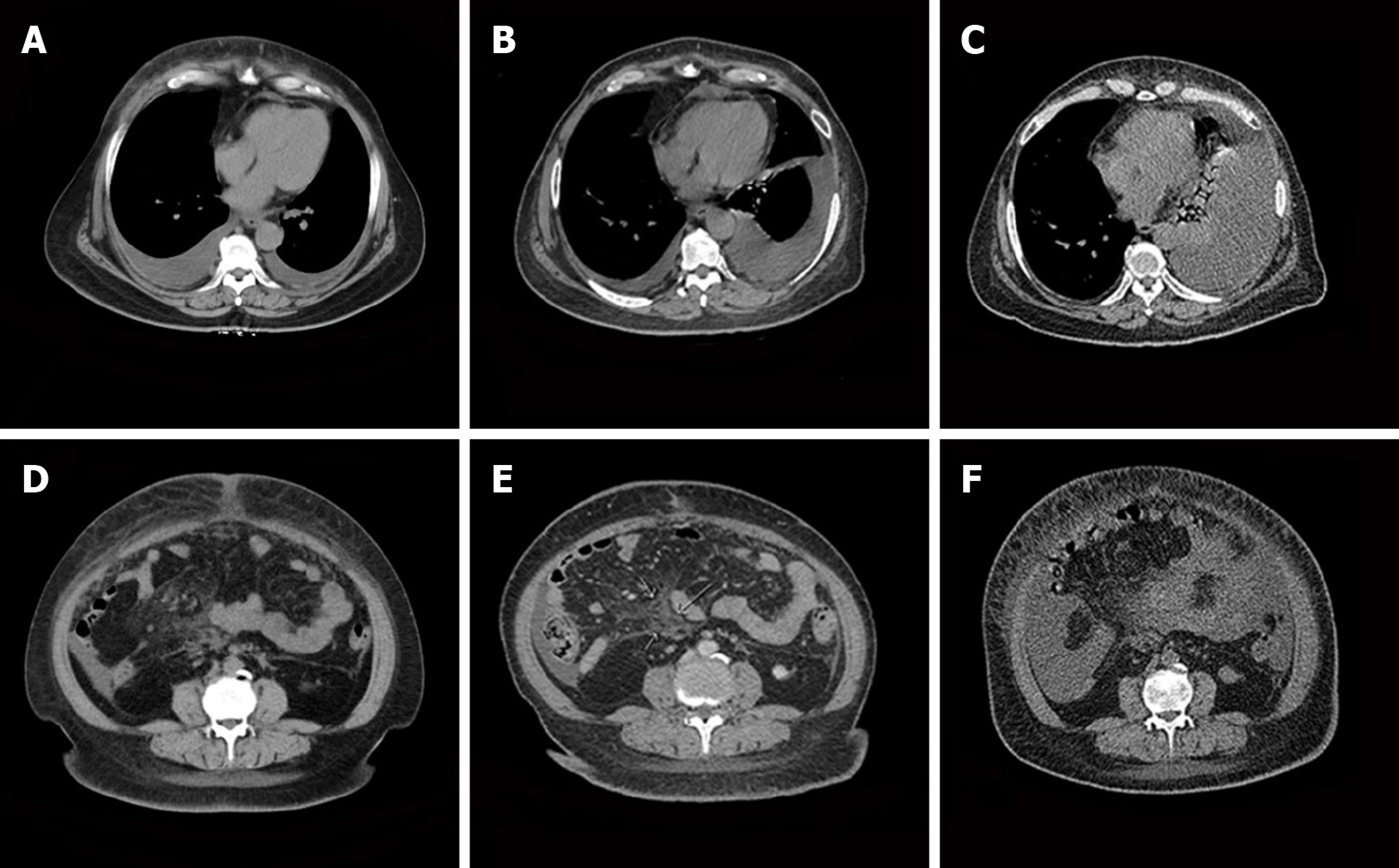
Figure 3 Computed tomography imaging of the patient's lesion.
A-C: Pleural effusion in the lungs was gradually increasing; D-F: Extensive diffuse lesions in the pelvic cavity.
- Citation: Ma YN, Bu HL, Jin CJ, Wang X, Zhang YZ, Zhang H. Peritoneal cancer after bilateral mastectomy, hysterectomy, and bilateral salpingo-oophorectomy with a poor prognosis: A case report and review of the literature. World J Clin Cases 2019; 7(22): 3872-3880
- URL: https://www.wjgnet.com/2307-8960/full/v7/i22/3872.htm
- DOI: https://dx.doi.org/10.12998/wjcc.v7.i22.3872


