
















©The Author(s) 2019.
World J Clin Cases. Nov 6, 2019; 7(21): 3524-3534
Published online Nov 6, 2019. doi: 10.12998/wjcc.v7.i21.3524
Published online Nov 6, 2019. doi: 10.12998/wjcc.v7.i21.3524
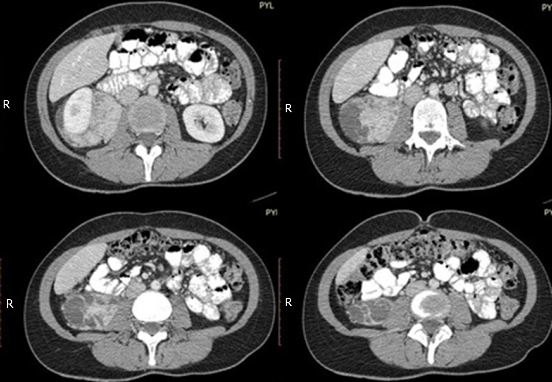
Figure 1 Abdominal computed tomography scan findings.
Selective coronal views of the abdominal computed tomography scan with intravenous and oral contrast material at 1.3 mm intervals. A solid, heterogeneous, retroperitoneal mass, anterior to the right psoas muscle, posterior to the right kidney, inferior to the right lobe of the liver, extending down to the level of the caecum, and pushing the right kidney anteriorly and laterally, is documented.
- Citation: Touloumis Z, Giannakou N, Sioros C, Trigka A, Cheilakea M, Dimitriou N, Griniatsos J. Retroperitoneal perivascular epithelioid cell tumours: A case report and review of literature. World J Clin Cases 2019; 7(21): 3524-3534
- URL: https://www.wjgnet.com/2307-8960/full/v7/i21/3524.htm
- DOI: https://dx.doi.org/10.12998/wjcc.v7.i21.3524