Copyright
©The Author(s) 2019.
World J Clin Cases. Oct 6, 2019; 7(19): 3126-3131
Published online Oct 6, 2019. doi: 10.12998/wjcc.v7.i19.3126
Published online Oct 6, 2019. doi: 10.12998/wjcc.v7.i19.3126
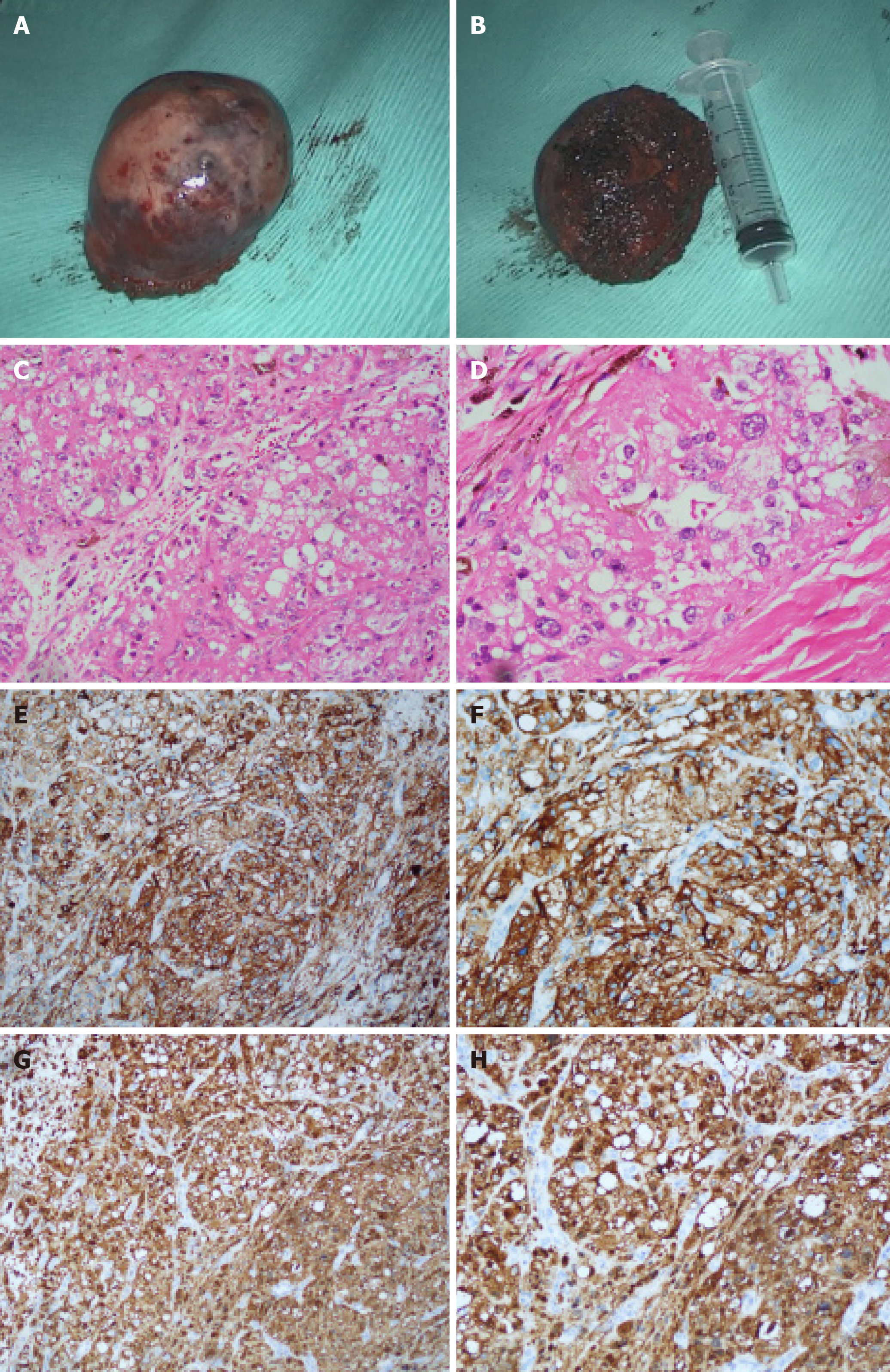
Figure 3 Cystic mass and immunohistochemical staining.
A, B: The tumor was removed under en bloc dissection with a relative clear margin; C, D: Hematoxylin-eosin stain: Neoplastic cells are arranged in irregular nests separated by fibrous septa. Cells are round or oval in shape with regular vesicular nuclei and prominent nucleoli, moderate to abundant eosinophilic or clear cytoplasm; E, F: Neoplastic cells showed a strong immunohistochemical expression of melanocytic marker: S100 (+); G, H: Neoplastic cells showed a strong immunohistochemical expression of melanocytic marker: HMB-45 (+).
- Citation: Chen YT, Yang Z, Li H, Ni CH. Clear cell sarcoma of soft tissue in pleural cavity: A case report. World J Clin Cases 2019; 7(19): 3126-3131
- URL: https://www.wjgnet.com/2307-8960/full/v7/i19/3126.htm
- DOI: https://dx.doi.org/10.12998/wjcc.v7.i19.3126


