Copyright
©The Author(s) 2019.
World J Clin Cases. Oct 6, 2019; 7(19): 3098-3103
Published online Oct 6, 2019. doi: 10.12998/wjcc.v7.i19.3098
Published online Oct 6, 2019. doi: 10.12998/wjcc.v7.i19.3098
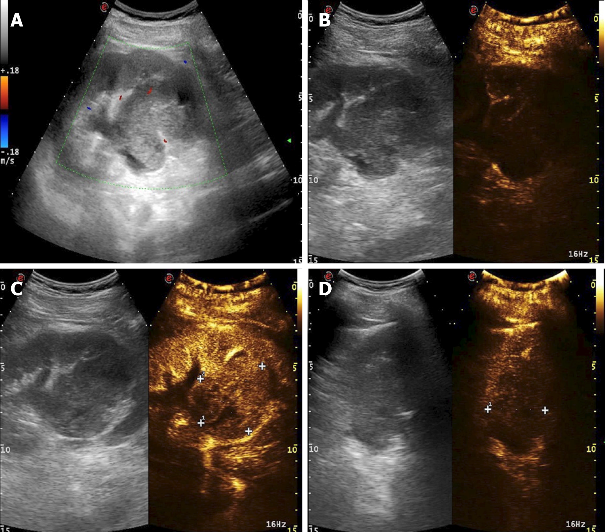
Figure 2 Contrast-enhanced ultrasound images of the mass.
A: Color Doppler flow imaging of the mass; B: The slow-moving performance; C: The hyperenhancement of the mass; D: The fast degeneration of the mass.
- Citation: Cai HJ, Cao N, Wang W, Kong FL, Sun XX, Huang B. Primary renal synovial sarcoma: A case report. World J Clin Cases 2019; 7(19): 3098-3103
- URL: https://www.wjgnet.com/2307-8960/full/v7/i19/3098.htm
- DOI: https://dx.doi.org/10.12998/wjcc.v7.i19.3098


