Copyright
©The Author(s) 2019.
World J Clin Cases. Oct 6, 2019; 7(19): 3039-3046
Published online Oct 6, 2019. doi: 10.12998/wjcc.v7.i19.3039
Published online Oct 6, 2019. doi: 10.12998/wjcc.v7.i19.3039
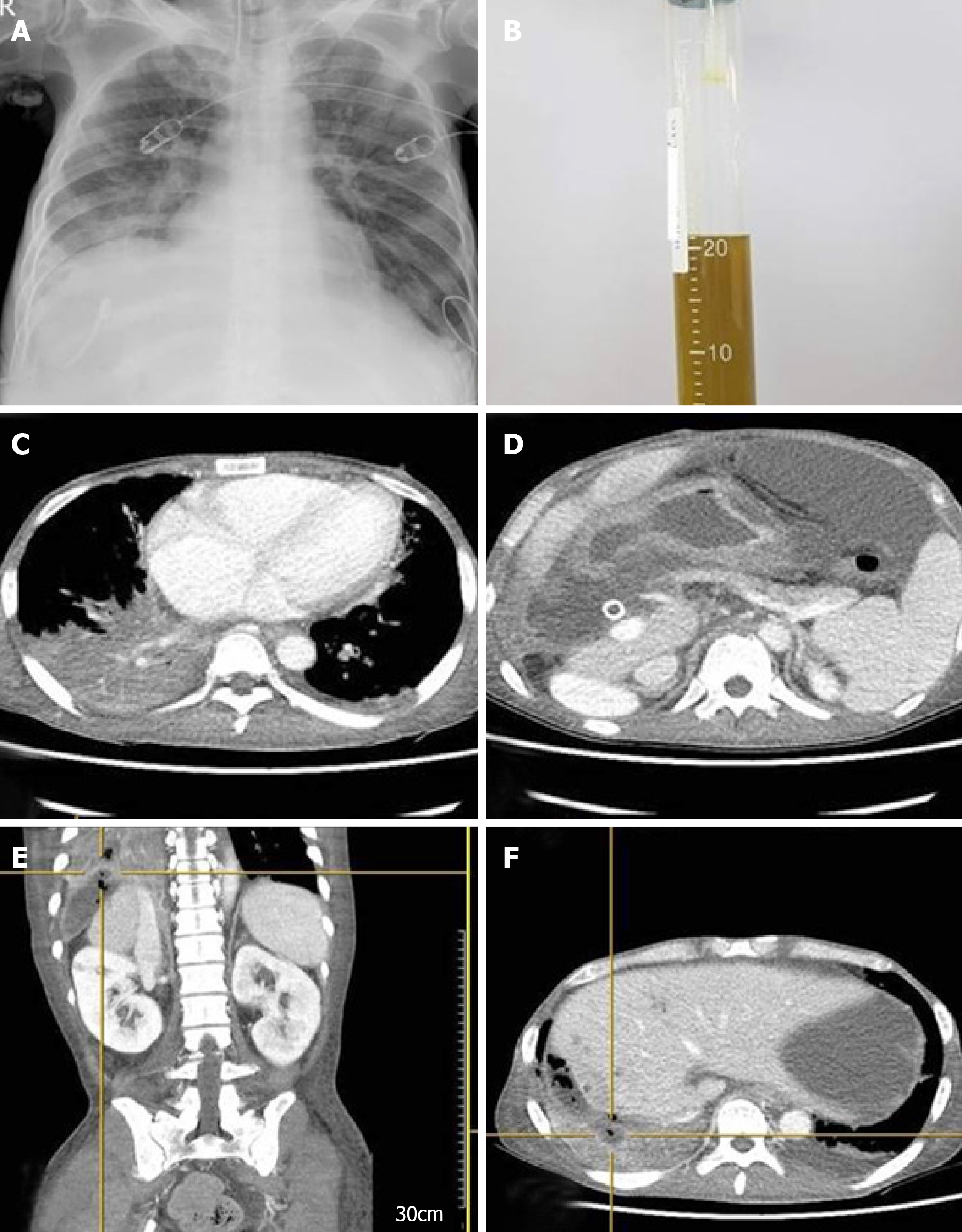
Figure 3 Imaging after emergency room due to respiratory faire.
A: Chest X-ray imaging after emergency room admission revealed pneumonic infiltration in the right lower lobe. B: Greenish bilious sputum was observed after intubation. C and D: Thorax and abdomen computed tomography (CT) showed pneumonic infiltration of the right lower lobe and free air in the perihepatic area, accompanied by fluid collection. E and F: Perforation of the diaphragm was also seen on CT, and bronchobiliary fistula was suspected.
- Citation: Kim HB, Na YS, Lee HJ, Park SG. Bronchobiliary fistula after ramucirumab treatment for advanced gastric cancer: A case report. World J Clin Cases 2019; 7(19): 3039-3046
- URL: https://www.wjgnet.com/2307-8960/full/v7/i19/3039.htm
- DOI: https://dx.doi.org/10.12998/wjcc.v7.i19.3039


