Copyright
©The Author(s) 2019.
World J Clin Cases. Oct 6, 2019; 7(19): 3039-3046
Published online Oct 6, 2019. doi: 10.12998/wjcc.v7.i19.3039
Published online Oct 6, 2019. doi: 10.12998/wjcc.v7.i19.3039
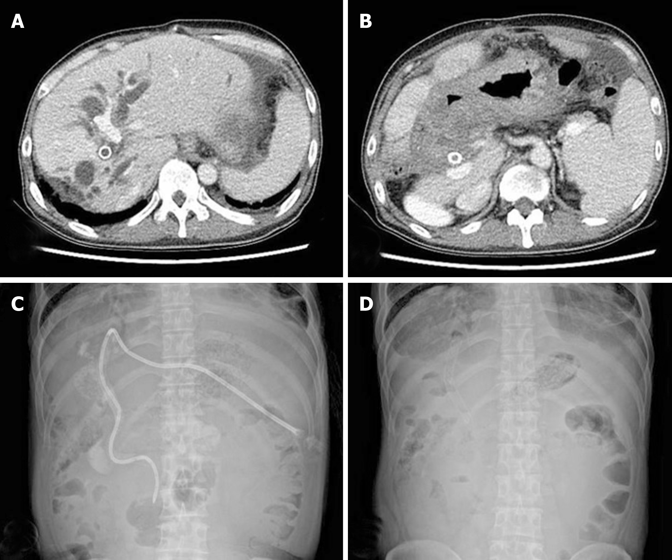
Figure 2 Imaging after first-line chemotherapy.
A and B: Computed tomography showed that the bile duct was dilated, indicating disease progression. C and D: To lower the elevated bilirubin level, percutaneous transhepatic biliary drainage and stent placement were performed again.
- Citation: Kim HB, Na YS, Lee HJ, Park SG. Bronchobiliary fistula after ramucirumab treatment for advanced gastric cancer: A case report. World J Clin Cases 2019; 7(19): 3039-3046
- URL: https://www.wjgnet.com/2307-8960/full/v7/i19/3039.htm
- DOI: https://dx.doi.org/10.12998/wjcc.v7.i19.3039


