Copyright
©The Author(s) 2019.
World J Clin Cases. Sep 26, 2019; 7(18): 2808-2814
Published online Sep 26, 2019. doi: 10.12998/wjcc.v7.i18.2808
Published online Sep 26, 2019. doi: 10.12998/wjcc.v7.i18.2808
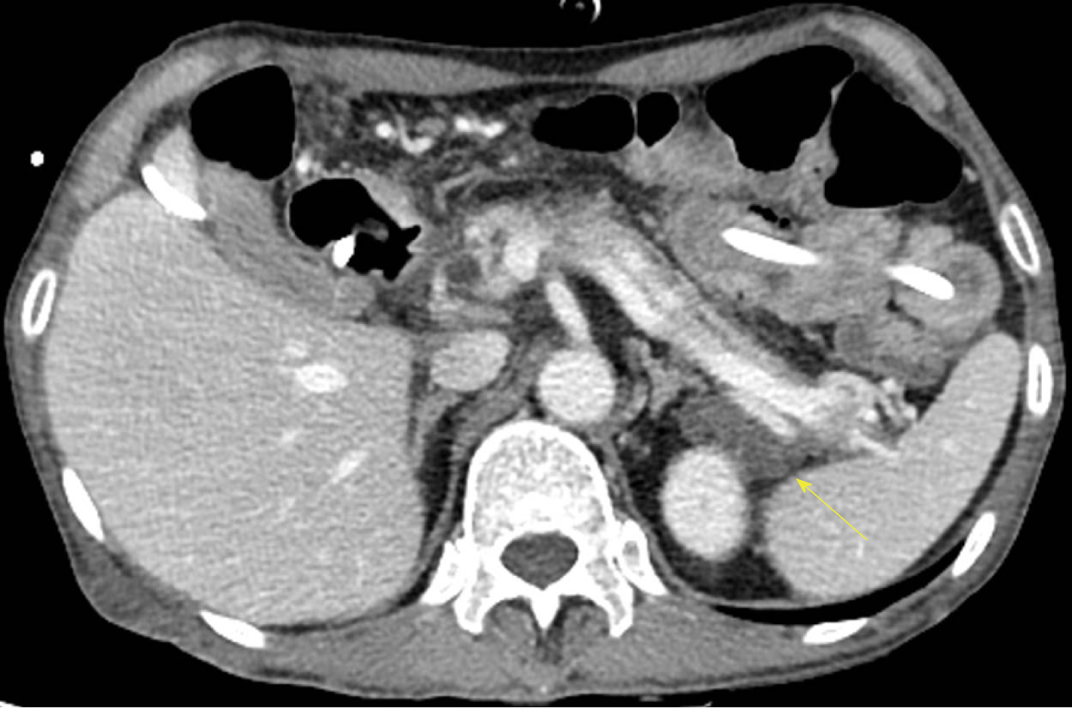
Figure 1 Computed tomography of the pancreas.
Cystic lesion with an attenuated lobulated margin along the peripancreatic tail and distal portion of the body of the pancreas (yellow arrow).
- Citation: Jo S, Song S. Pancreatitis, panniculitis, and polyarthritis syndrome caused by pancreatic pseudocyst: A case report. World J Clin Cases 2019; 7(18): 2808-2814
- URL: https://www.wjgnet.com/2307-8960/full/v7/i18/2808.htm
- DOI: https://dx.doi.org/10.12998/wjcc.v7.i18.2808


