Copyright
©The Author(s) 2019.
World J Clin Cases. Sep 6, 2019; 7(17): 2617-2622
Published online Sep 6, 2019. doi: 10.12998/wjcc.v7.i17.2617
Published online Sep 6, 2019. doi: 10.12998/wjcc.v7.i17.2617
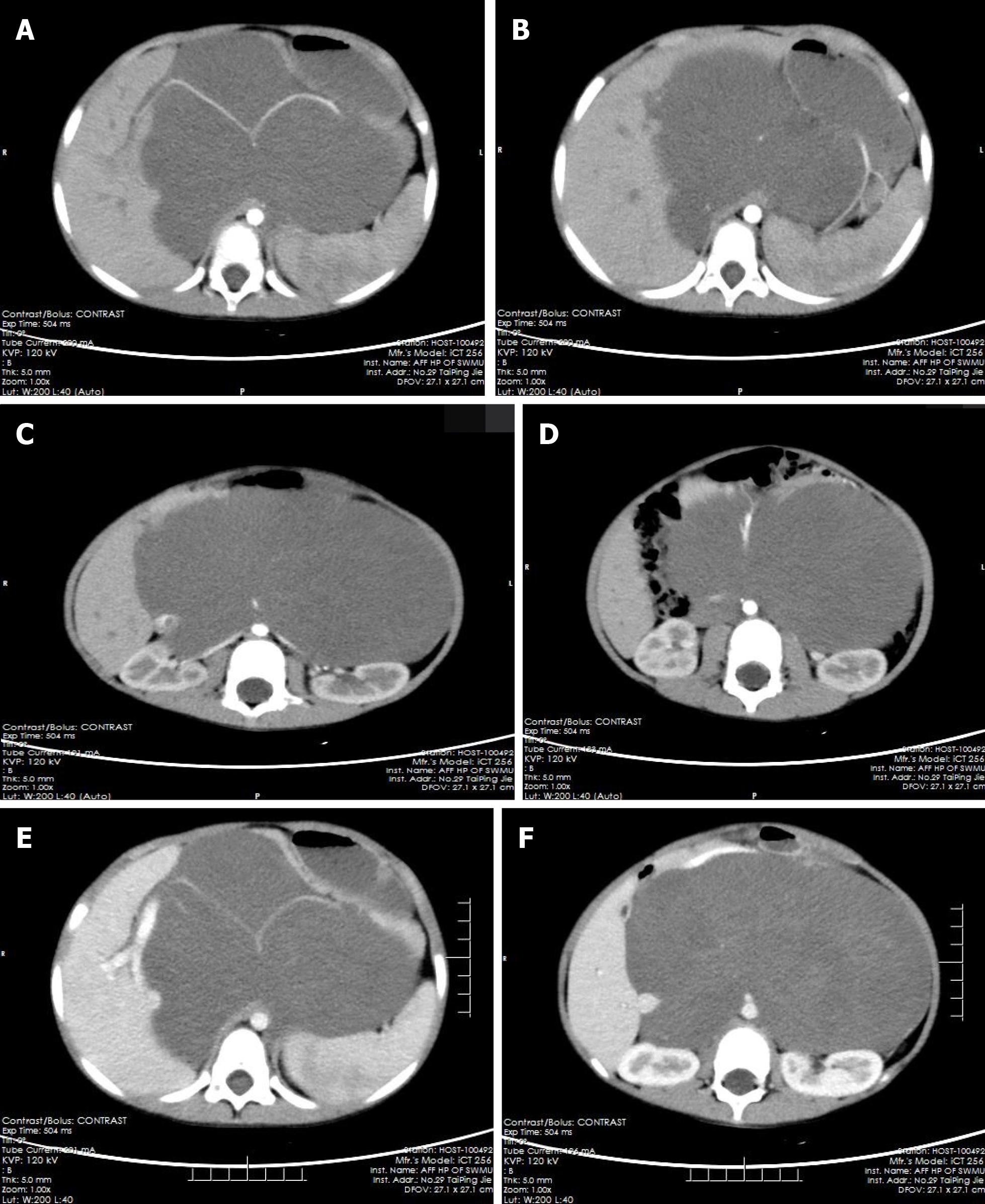
Figure 2 The computerized tomographic images of tumors surrounding blood vessels.
A-D: Tumor-enclosed arteries: hepatic artery (A), splenic artery (A, B), bilateral renal artery (C), superior mesenteric artery (D); E, F: Tumor-enclosed veins: Portal vein (E) and inferior vena cava (F).
- Citation: Zheng X, Luo L, Han FG. Cause of postprandial vomiting - a giant retroperitoneal ganglioneuroma enclosing large blood vessels: A case report. World J Clin Cases 2019; 7(17): 2617-2622
- URL: https://www.wjgnet.com/2307-8960/full/v7/i17/2617.htm
- DOI: https://dx.doi.org/10.12998/wjcc.v7.i17.2617


