Copyright
©The Author(s) 2019.
World J Clin Cases. Sep 6, 2019; 7(17): 2580-2586
Published online Sep 6, 2019. doi: 10.12998/wjcc.v7.i17.2580
Published online Sep 6, 2019. doi: 10.12998/wjcc.v7.i17.2580
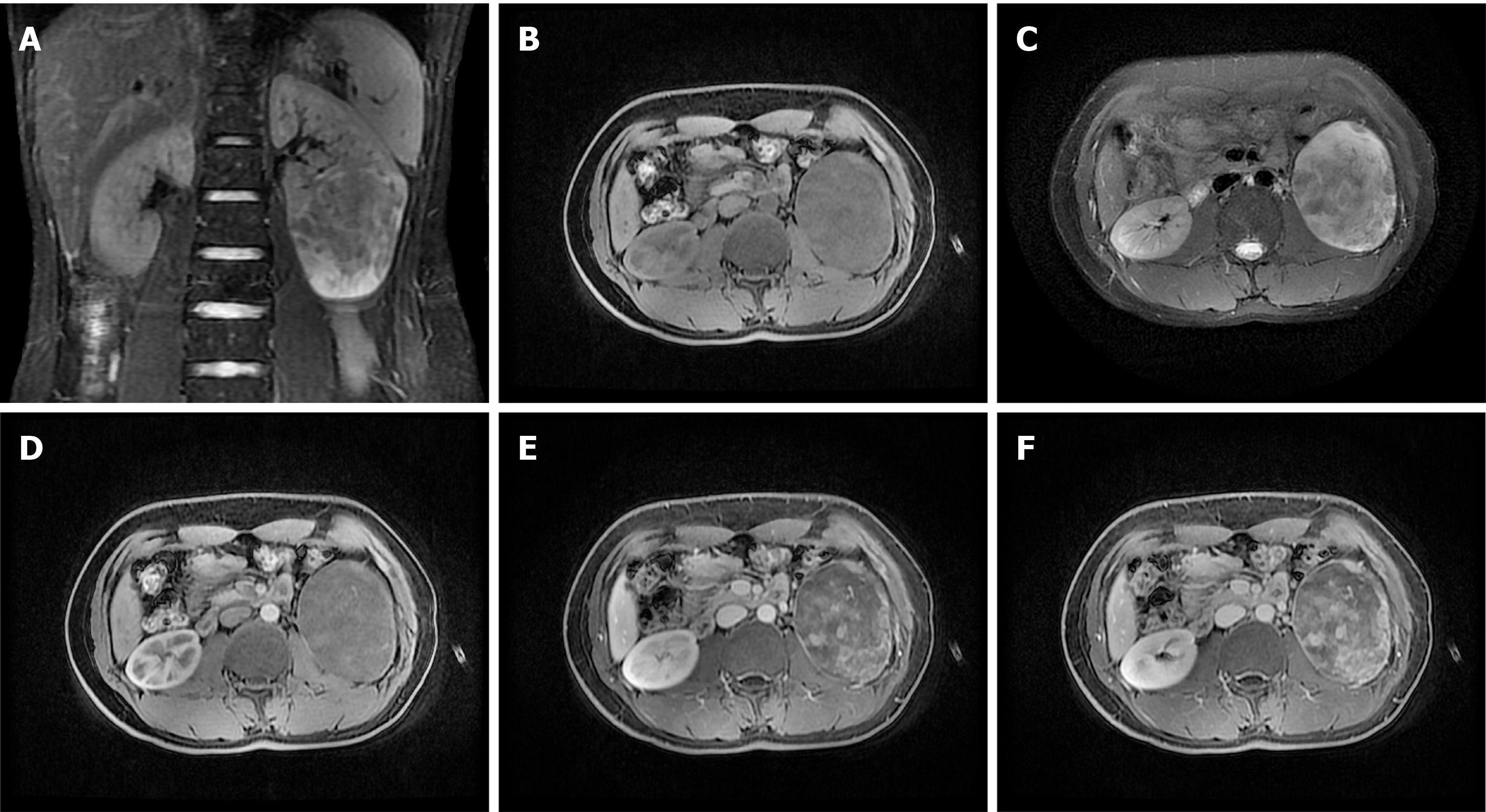
Figure 2 Moderate or mild enhancement in the solid component after contrast enhancement.
A: Coronal T2-weighted image showing an exophytic cystic-solid mass in the lower pole of the left kidney; B: Cystic region shows T1 hypointensity and T2 hyperintensity; C: Solid region shows T1 hypointensity and T2 hyperintensity; D: Contrast-enhanced magnetic resonance imaging during the corticomedullary phase; E: Nephrographic phase; F: Excretory phase showing that the tumor’s cystic parts were not enhanced and the tumor’s solid parts were enhanced to a moderate or mild degree that increased with time.
- Citation: Ye J, Xu Q, Zheng J, Wang SA, Wu YW, Cai JH, Yuan H. Imaging of mixed epithelial and stromal tumor of the kidney: A case report and review of the literature. World J Clin Cases 2019; 7(17): 2580-2586
- URL: https://www.wjgnet.com/2307-8960/full/v7/i17/2580.htm
- DOI: https://dx.doi.org/10.12998/wjcc.v7.i17.2580


