
















©The Author(s) 2019.
World J Clin Cases. Aug 6, 2019; 7(15): 2128-2133
Published online Aug 6, 2019. doi: 10.12998/wjcc.v7.i15.2128
Published online Aug 6, 2019. doi: 10.12998/wjcc.v7.i15.2128
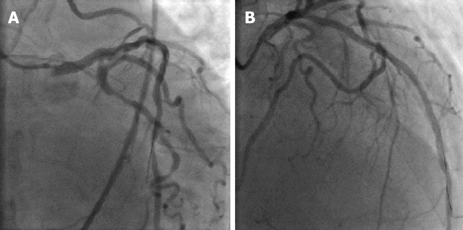
Figure 3 Coronary angiograms after percutaneous coronary intervention.
A: Caudal view; B: Right anterior oblique cranial view. The images show excellent stent expansion and no dissection in the left main coronary artery ostia, the mid segment of left anterior descending artery, and right coronary artery with thrombolysis in myocardial infarction grade 3 flow.
- Citation: Wu Q, Li ZZ, Yue F, Wei F, Zhang CY. Percutaneous coronary intervention for ostial lesions of the left main stem in a patient with congenital single left coronary artery: A case report. World J Clin Cases 2019; 7(15): 2128-2133
- URL: https://www.wjgnet.com/2307-8960/full/v7/i15/2128.htm
- DOI: https://dx.doi.org/10.12998/wjcc.v7.i15.2128