Copyright
©The Author(s) 2019.
World J Clin Cases. Aug 6, 2019; 7(15): 2110-2119
Published online Aug 6, 2019. doi: 10.12998/wjcc.v7.i15.2110
Published online Aug 6, 2019. doi: 10.12998/wjcc.v7.i15.2110
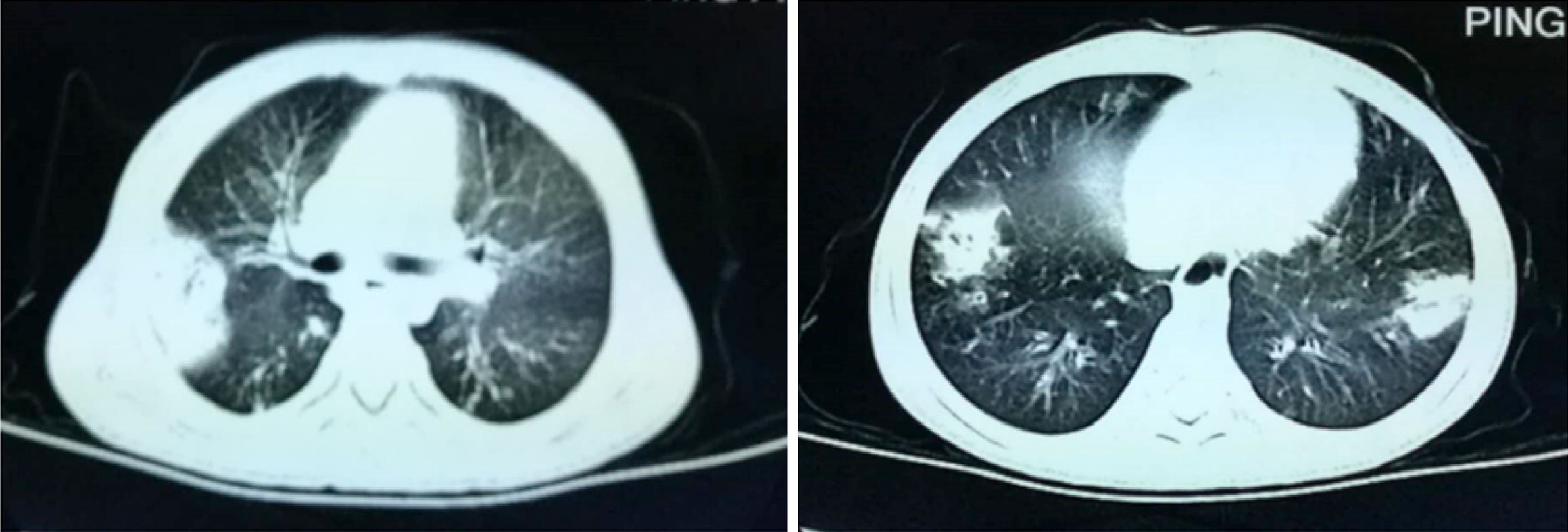
Figure 1 Chest computed tomography images of the cystic fibrosis patient.
A chest computed tomography scan showed obvious exudative lesions and bilateral bronchiectasis in the lung of the cystic fibrosis patient.
- Citation: Wang YQ, Hao CL, Jiang WJ, Lu YH, Sun HQ, Gao CY, Wu M. c.753_754delAG, a novel CFTR mutation found in a Chinese patient with cystic fibrosis: A case report and review of the literature. World J Clin Cases 2019; 7(15): 2110-2119
- URL: https://www.wjgnet.com/2307-8960/full/v7/i15/2110.htm
- DOI: https://dx.doi.org/10.12998/wjcc.v7.i15.2110


