Copyright
©The Author(s) 2019.
World J Clin Cases. May 26, 2019; 7(10): 1234-1241
Published online May 26, 2019. doi: 10.12998/wjcc.v7.i10.1234
Published online May 26, 2019. doi: 10.12998/wjcc.v7.i10.1234
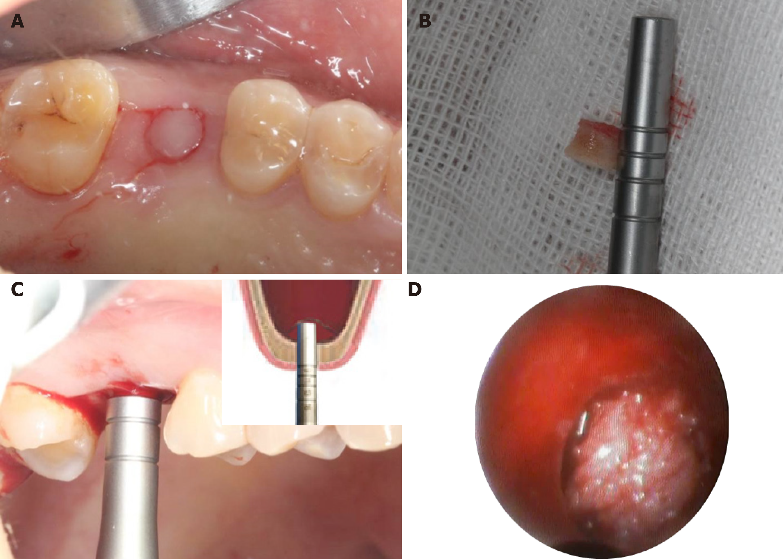
Figure 2 Intraoperative illustrations to explain each step of the surgery.
A: Establishment of a punch incision; B: Full-thickness keratinized gingival punch removal; C: Gentle elevation of the Schneiderian membrane till approximately 7 mm; D: An endoscope was used to monitor the maxillary sinus membrane.
- Citation: Mudalal M, Sun XL, Li X, Fang J, Qi ML, Wang J, Du LY, Zhou YM. Minimally invasive endoscopic maxillary sinus lifting and immediate implant placement: A case report. World J Clin Cases 2019; 7(10): 1234-1241
- URL: https://www.wjgnet.com/2307-8960/full/v7/i10/1234.htm
- DOI: https://dx.doi.org/10.12998/wjcc.v7.i10.1234


